586 - Timeliness of First Neuroimaging for Suspected Stroke in a Pediatric Emergency Department
Friday, April 24, 2026
5:30pm - 8:00pm ET
Publication Number: 1563.586
Lynn Babcock, Cincinnati Children's Hospital Medical Center, Cincinnati, OH, United States; Kate E. Lex, Cincinnati Children's Hospital Medical Center, Cincinnati, OH, United States; Natalie Cimpello, Cincinnati Children's Hospital Medical Center, Cincinnati, OH, United States; Ryan Murphy, Cincinnati Children's Hospital Medical Center, Cincinnati, OH, United States; Chen Chen, Cincinnati Children's Hospital Medical Center, Basking Ridge, NJ, United States; Benjamin T. Kerrey, Cincinnati Children's Hospital Medical Center, Cincinnati, OH, United States; J. Michael Taylor, Cincinnati Children's Hospital Medical Center, Cincinnati, OH, United States; Mary E. Frey, ENA Cincinnati Children's Hosptial Medical Center, Cincinnati, OH, United States; Karen Ahaus, Cincinnati Children's Hospital, Sunman, IN, United States; James M. Gray, Cincinnati Children's Hospital Medical Center, Cincinnati, OH, United States
Professor, Department of Pediatrics, Division of Pediatric Emergency Medicine Cincinnati Children's Hospital Medical Center Cincinnati, Ohio, United States
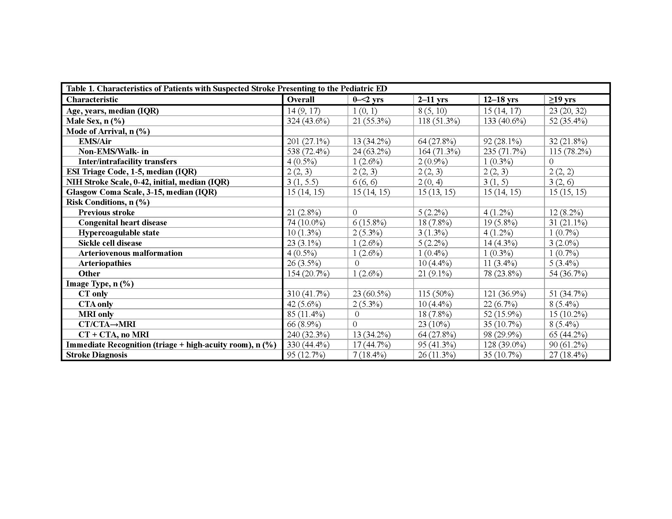
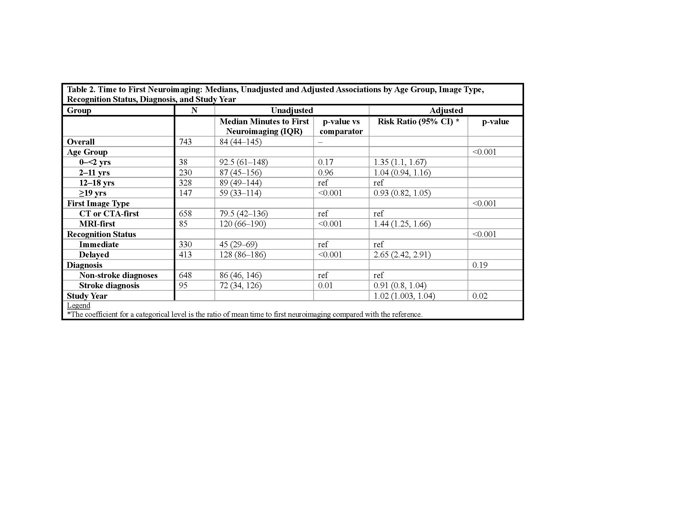
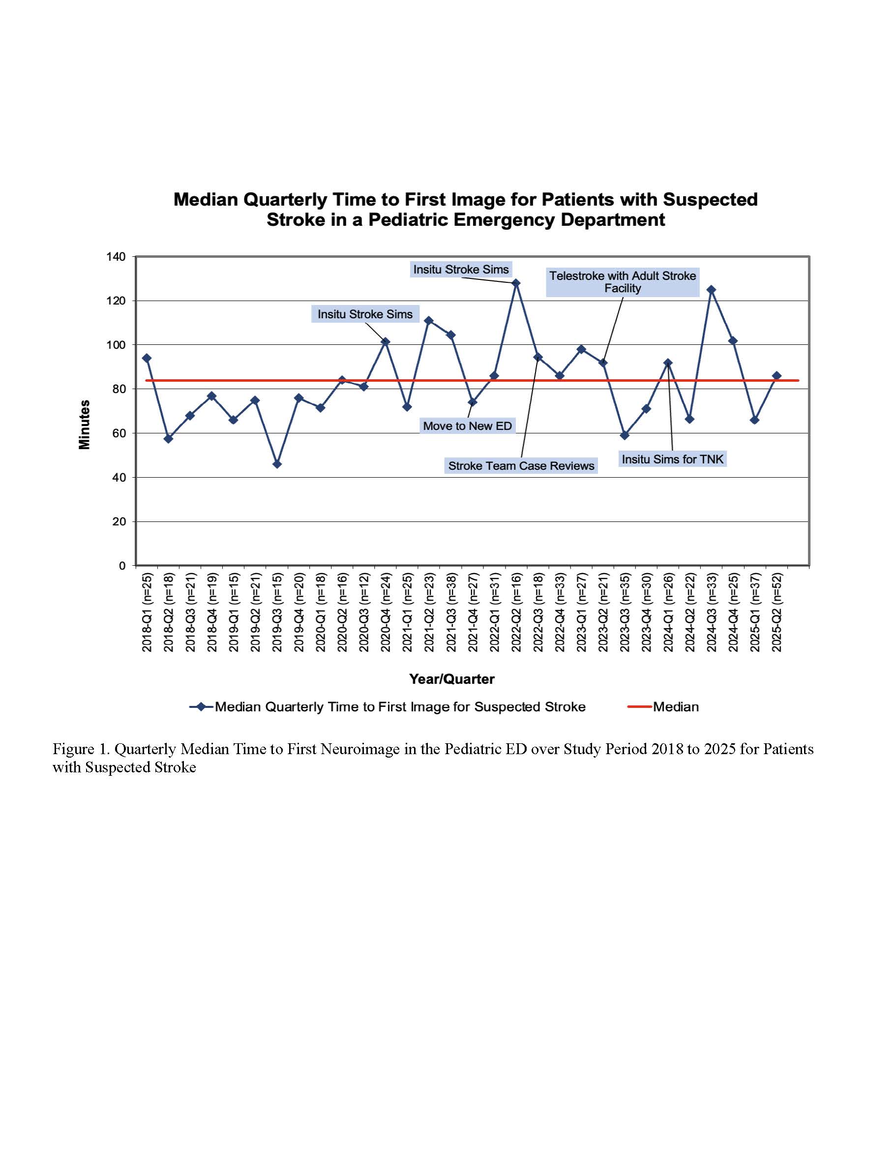
Background: Timely neuroimaging is essential for accurate stroke diagnosis in the emergency department (ED). In children, variation in age, imaging practices, stroke risk recognition may contribute to delays, yet pediatric ED data are limited. Objective: To characterize patients evaluated for suspected stroke in a pediatric ED, assess the timeliness of first neuroimaging by age, image modality, recognition timing, and diagnosis, and examine temporal trends. Design/Methods: Retrospective cohort of patients with suspected stroke who underwent neuroimaging in a pediatric ED from January 1, 2018 through June 30, 2025. Time to first image was measured from ED arrival to scan start using electronic health record timestamps. Timeliness was compared across age group (0- < 2, 2-11, 12-18, ≥19 years), image modality (CT/CTA-first vs MRI-first), timing of stroke-risk recognition (immediate, defined as triage nurse suspicion with placement in a high-acuity area; vs delayed), and diagnosis (confirmed stroke vs non-stroke). Multivariable linear regression of log-transformed time to first image assessed independent predictors, adjusting for age group, modality, recognition timing, confirmed stroke status, and study year. Quarterly median times to first image were plotted on a run chart. Temporal trends of time to first image (log-transformed) were assessed with longitudinal log-linear models including calendar year, group, and yearXgroup, and effect of time was tested for each level of group. Results: Among 743 patients, 95 (12.7%) had confirmed stroke (65 ischemic, 15 hemorrhagic, 11 TIA, 4 mixed). Median age was 14 years (IQR 9-17); 43.6% were male [Table 1]. Median time to first neuroimaging was 84 minutes (IQR 45-145) [Figure 1]. MRI-first rose from 8.4% (2018) to 14.6% (2025). Younger children had the longest times (92.5 min) vs 12-18 and ≥19 y (89 and 59 min; p<.001) MRI-first was slower than CT-first (120 vs 79.5 min; p<.001). Imaging was faster for those with confirmed stroke versus non-stroke (72 vs 86 min; p=.01). In adjusted models, younger age, MRI-first imaging, and delayed-recognition were independently associated with longer time to first image (all p<.001) [Table 2]. Across years, time to first image was marginally longer (p=.02); times were stable by age and modality, rose in delayed-recognition (p=.002), and decreased in confirmed stroke (p=.004).
Conclusion(s): Younger age, MRI-first imaging, and delayed recognition were associated with longer time to first neuroimaging, while timeliness improved modestly over the study period. Earlier recognition and streamlined pathways may reduce pediatric stroke imaging delays.
Table 1. Characteristics of Patients with Suspected Stroke Presenting to the Pediatric ED
Table 2. Time to First Neuroimaging: Medians, Unadjusted and Adjusted Associations by Age Group, Image Type, Recognition Status, Diagnosis, and Study Year
Figure 1. Quarterly Median Time to First Neuroimage in the Pediatric ED over Study Period 2018 to 2025 for Patients with Suspected Stroke