
Neonatal Neurology
Session: Neonatal Neurology 3: Clinical - Term 3
.jpg "Rachel L. Leon, MD, PhD (she/her/hers) photo")
Rachel L. Leon, MD, PhD (she/her/hers)
Assistant Professor of Pediatrics
University of Texas Southwestern Medical School
Dallas, Texas, United States
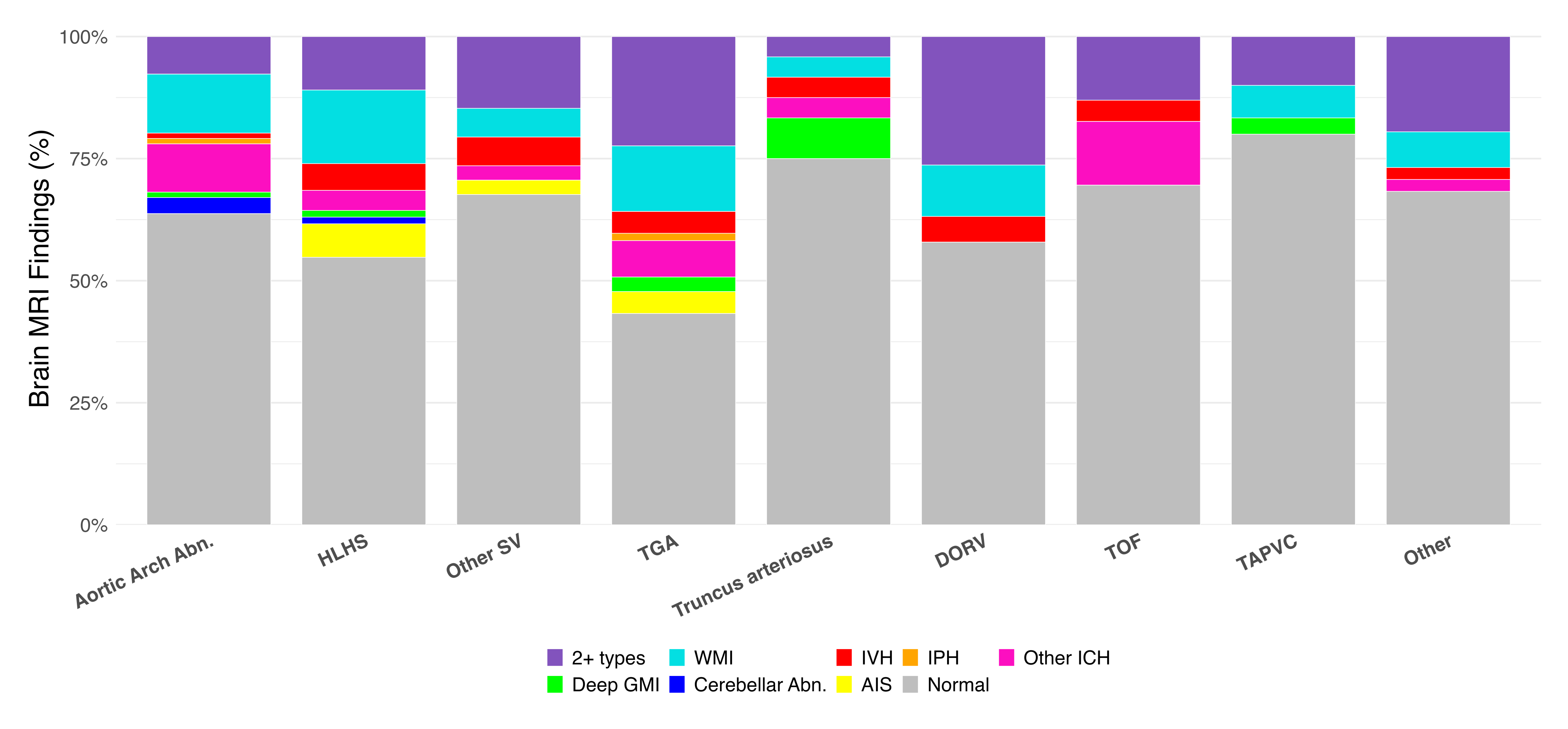 Stacked bar graph of the prevalence of specific types of brain MRI findings in each diagnostic category of CHD. HLHS: hypoplastic left heart syndrome; SV: single ventricle physiology; TGA: transposition of the great arteries; DORV: double-outlet right ventricle; TOF: tetralogy of Fallot; TAPVC: total anomalous pulmonary venous connection; WMI: white matter injury; IVH: intraventricular hemorrhage; IPH: intraparenchymal hemorrhage; ICH: intracranial hemorrhage; GMI: gray matter injury; AIS: arterial ischemic stroke
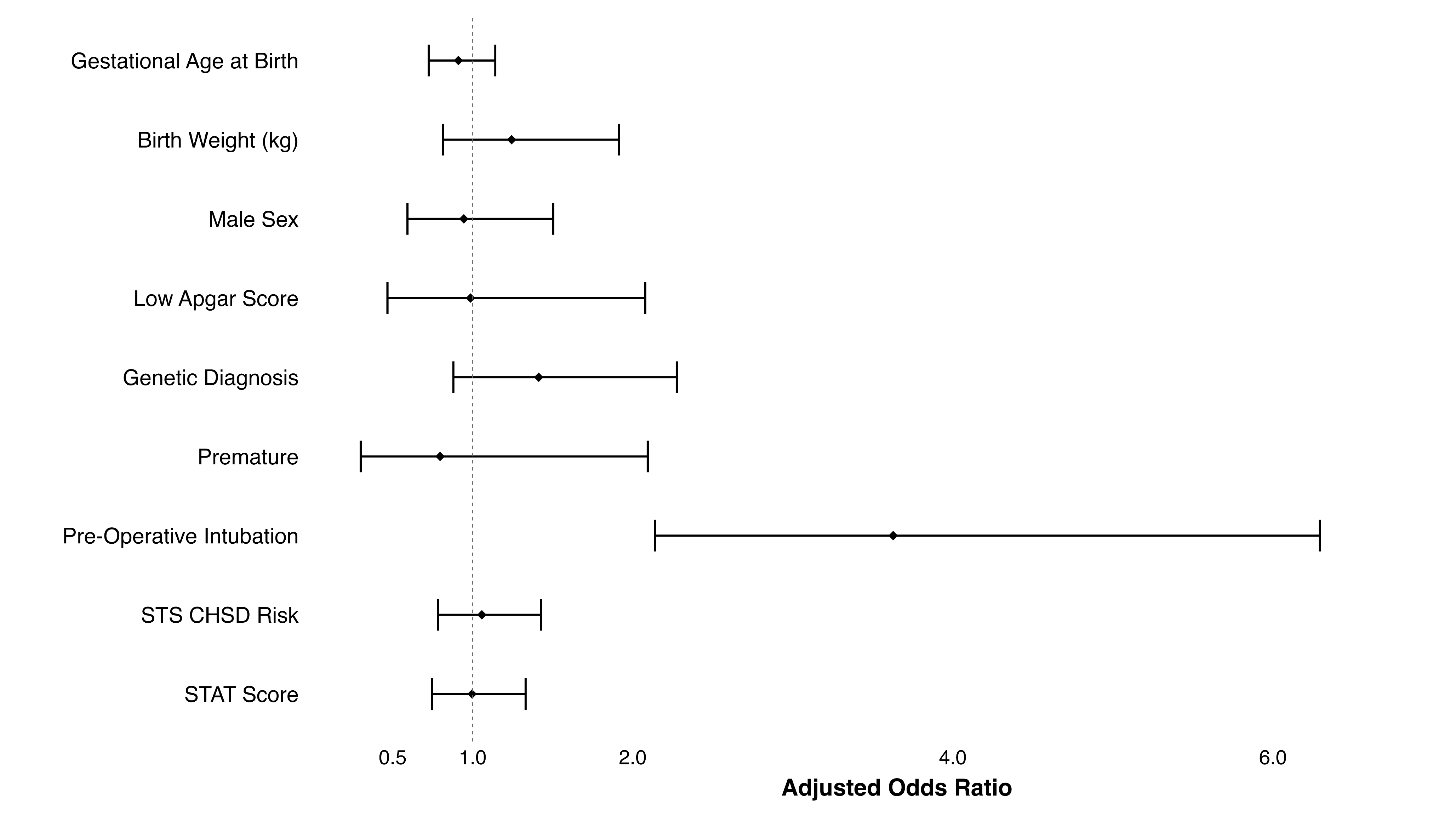 Forest plot showing adjusted odds ratios (aORs) and 95% confidence intervals (CIs) from a multivariable logistic regression model evaluating associations between clinical characteristics and abnormal pre-operative brain MRI findings. Predictors used in the model included gestational age at birth (weeks), birth weight (kg),male sex, low Apgar score (defined as an Apgar score <6 at either 1 or 5 minutes), presence of a genetic diagnosis, prematurity (gestational age <37 weeks), pre-operative intubation, assigned STS CHSD Risk, and STAT surgical complexity score. The dashed vertical line represents an aOR of 1 indicating no association. Pre-operative intubation was the only variable significantly associated with abnormal brain MRI findings (aOR = 3.5, 95% CI 2.1-6.3, p<0.0001).
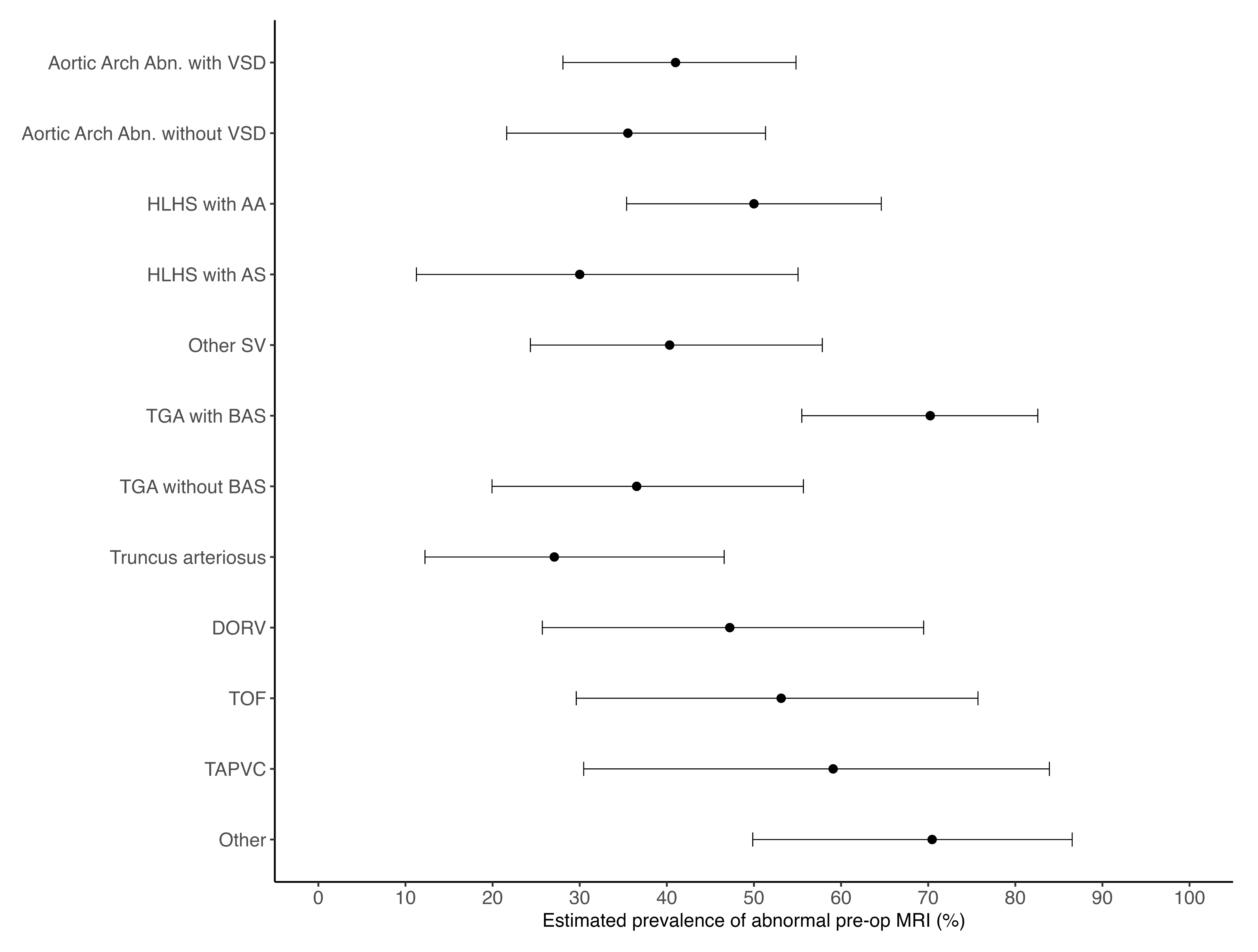 Firth-penalized logistic regression without an intercept (one estimate per group; no reference group) showing estimated prevalence of abnormal pre-operative brain MRI with 95% confidence intervals in each of the diagnostic subgroups of CHDs in the cohort. VSD: ventricular septal defect; AA: aortic atresia; AS: aortic stenosis; SV: single ventricle physiology; BAS: balloon atrial septostomy; DORV: double outlet right ventricle; TOF: tetralogy of Fallot; TAPVC: total anomalous pulmonary venous connection.