57 - Hypocapnic Episodes Are Not Associated With Abnormal MRI Findings in Neonates With Hypoxic-Ischemic Encephalopathy Treated With Therapeutic Hypothermia
Friday, April 24, 2026
5:30pm - 8:00pm ET
Publication Number: 1050.57
Junkai Wen, University of Alabama School of Medicine, Birmingham, AL, United States; Tao Chen, UAB, Spanish Fort, AL, United States; Namasivayam Ambalavanan, University of Alabama School of Medicine, Birmingham, AL, United States; Vivek V. Shukla, University of Alabama at Birmingham, Birmingham, AL, United States
Fellow University of Alabama School of Medicine Birmingham, Alabama, United States
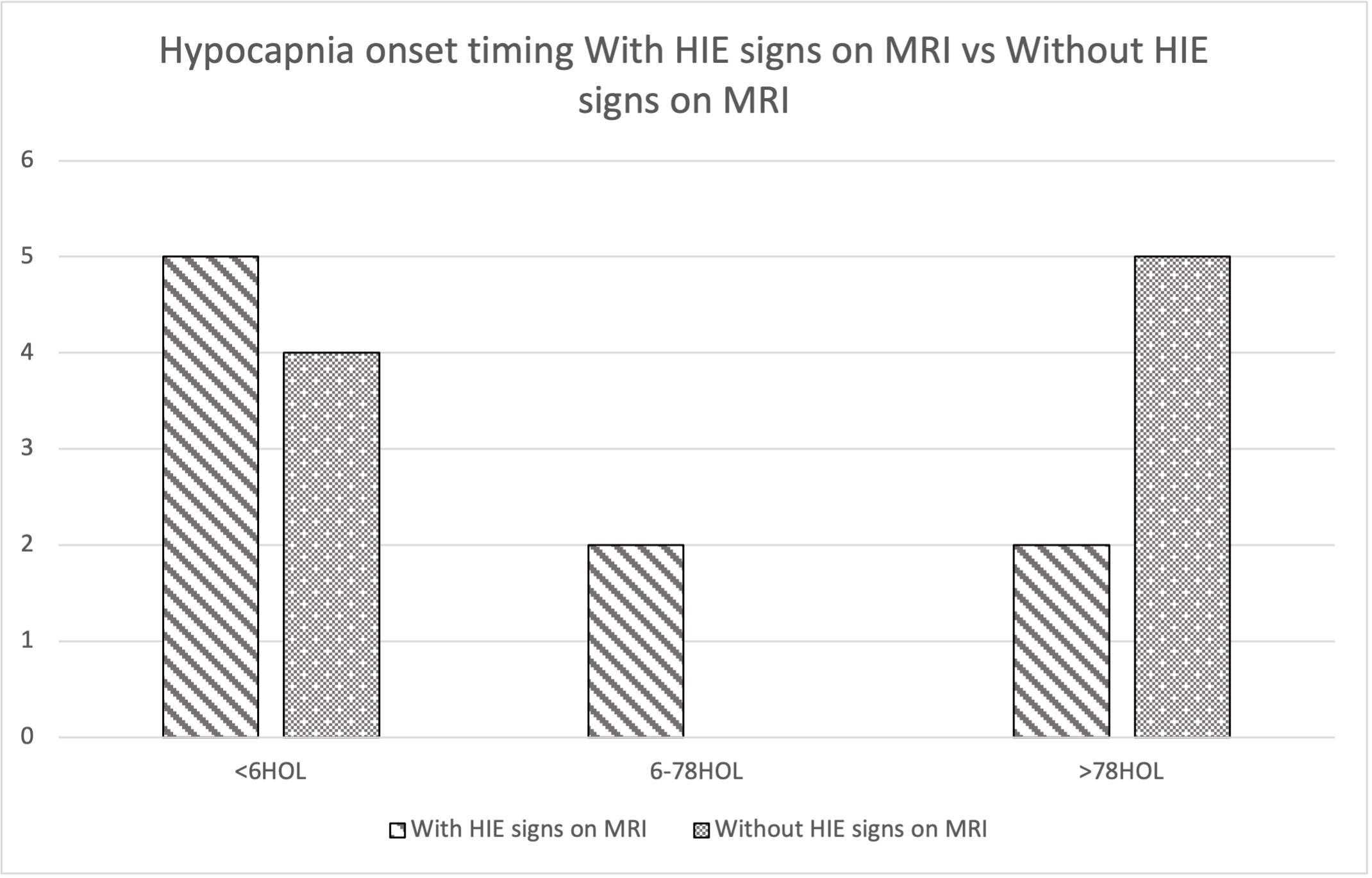
Background: In neonates, cerebral blood flow is sensitive to fluctuations in carbon dioxide(CO2) levels. Hypo-capnic episodes can lead to cerebral vasoconstriction. Previous studies have shown that hypo-capnia is associated with an increased risk of cerebral palsy and neuro-developmental impairment in preterm infants. Previous studies in term infants with hypoxic-ischemic encephalopathy(HIE) have shown unclear association between early hypo-capnia and adverse neurological outcomes, with some studies suggesting a positive association and others found no significant link. Objective: This study aims to explore whether episodes of hypo-capnia, as detected by intermittent blood gas analysis in the early postnatal period, are associated with abnormal brain MRI findings in neonates with HIE treated with therapeutic hypothermia. Design/Methods: This single-center, retrospective cohort study included 61 neonates with HIE who underwent therapeutic hypothermia in the regional neonatal ICU at University of Alabama At Birmingham from 2021 to 2025. Abnormal CO2 values within the first 100 hours of life were classified as: Severe hypo-capnia: pCO2 ≤ 19.5 mmHg, Moderate hypo-capnia: 19.5 mmHg < pCO2 ≤ 24.8 mmHg Among the cohort, 10 infants had severe hypo-capnic episodes, 16 had moderate hypo-capnic episodes. Among them, 4 infants had both severe and moderate hypo-capnic episodes. Abnormal hypo-capnic episodes were grouped as early ( < 6 hours of life), mid (6-78 hours), or late (>78 hours), based on blood gas sampling time. Brain injury was assessed by MRI performed within 3 days after rewarming. Associations between abnormal hypo-capnic episodes and adverse outcomes (HIE signs on brain MRI) were analyzed using chi-square tests and binary logistic regression. Results: A total of 684 blood gas results from 61 neonates were collected. Of the 61 neonates, 25 had brain MRI signs of HIE, 35 had normal MRIs, and 1 had no recorded MRI. Hypocapnic episodes occurred in 26 neonates, with 14 having HIE on MRI and 12 having normal MRIs. Severe hypocapnic episodes were observed in 6 infants with HIE and 4 without, while moderate episodes occurred in 8 infants with HIE and 8 without. Chi-square and binary logistic regression tests revealed no statistically significant associations between severe or moderate hypocapnia and HIE on MRI.
Conclusion(s): No direct associations were observed between hypo-capnic episodes and adverse MRI findings in this cohort.
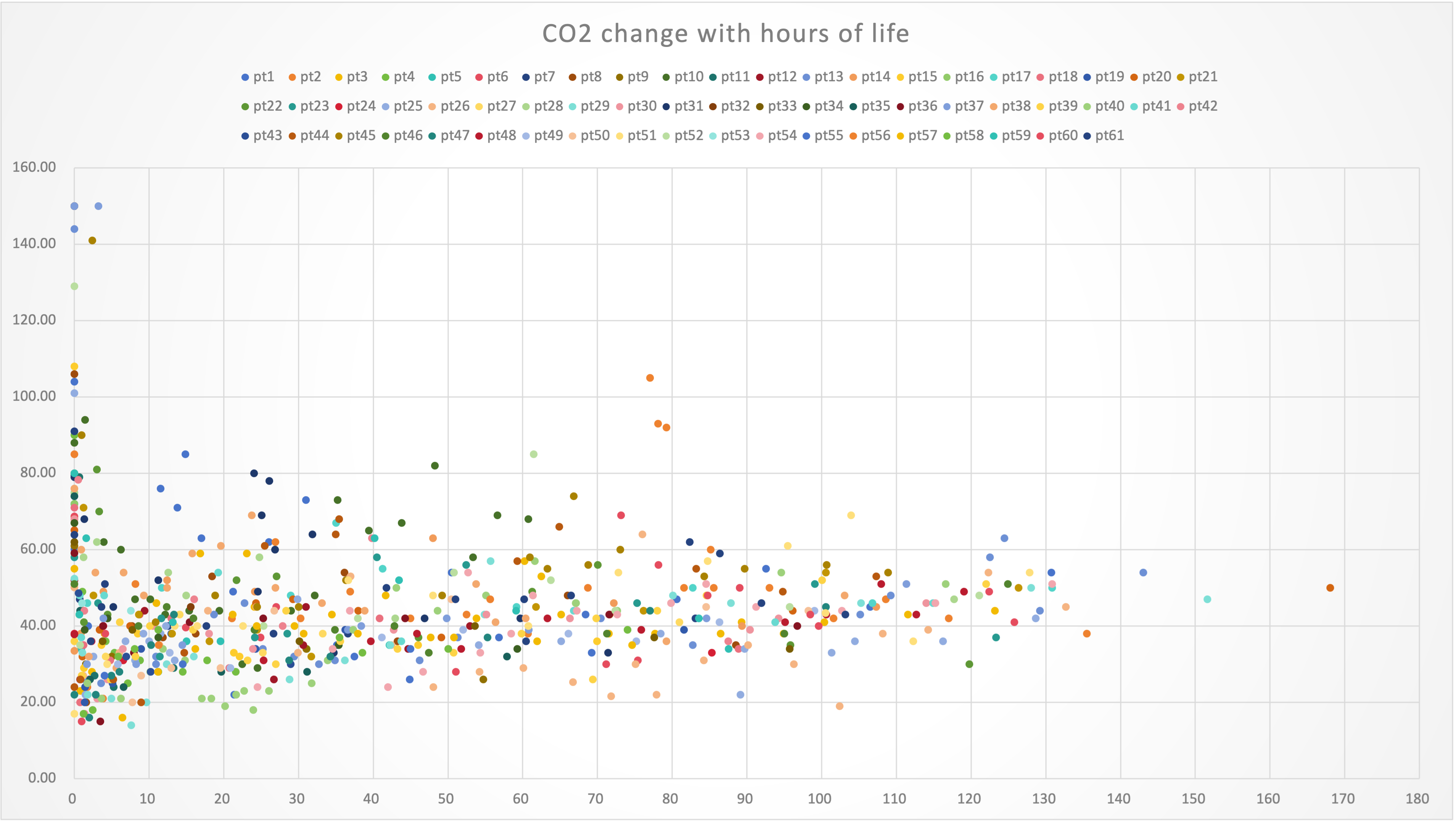
CO2 level distribution with hours of life
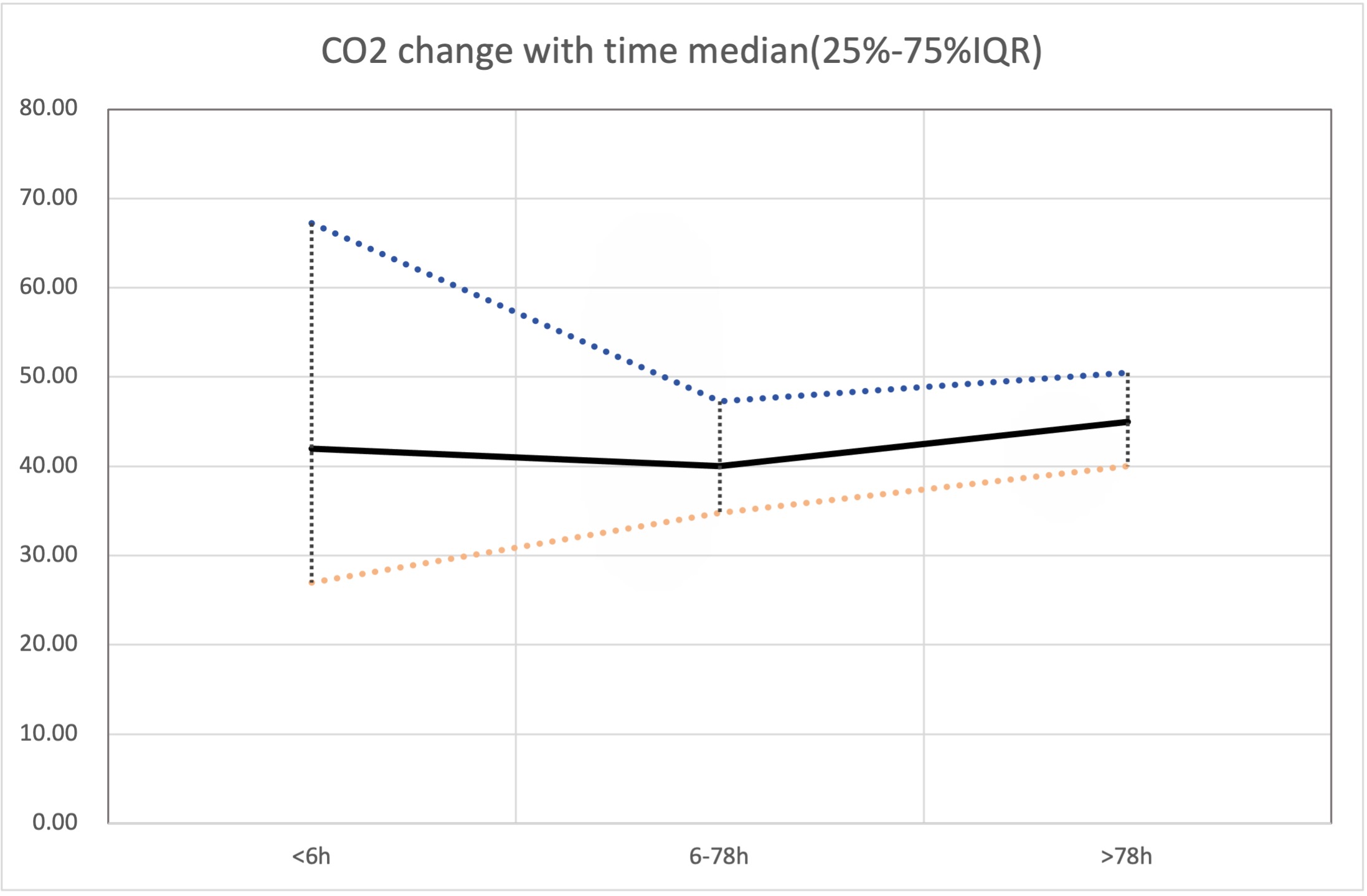
CO2 level distribution in 3 different time slots median with 25%/75% IQR
Early vs Mid vs Late onset of hypocapnia MRI with HIE signs vs MRI without HIE signs

 photo")