
Neonatal Neurology
Session: Neonatal Neurology 6: Prenatal

Jung-Hoon Kim, PhD
Research Faculty
Children's National Health System
Washington, District of Columbia, United States
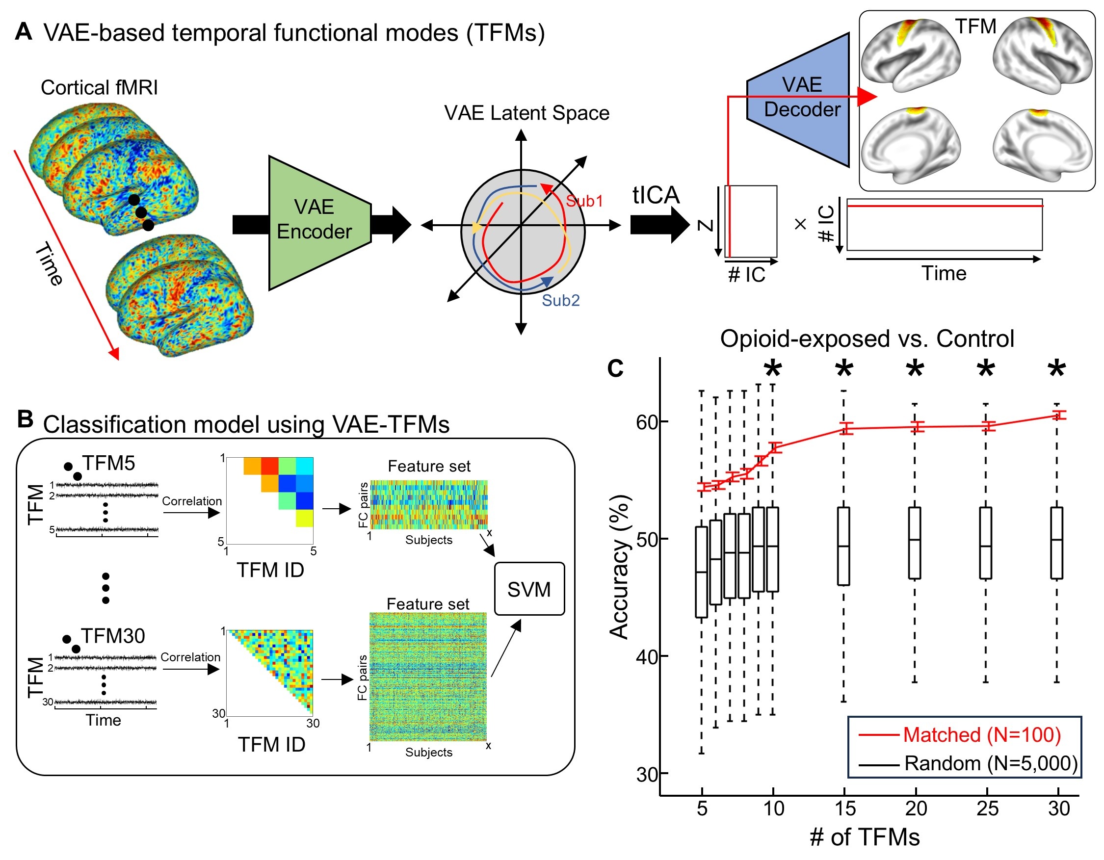 (A) Cortical resting-state fMRI patterns are encoded into a latent space via a VAE, generating latent trajectories that capture nonlinear temporal dynamics. Temporal independent component analysis (tICA) is then applied within this latent space to extract TFMs, which are subsequently projected back onto the cortical surface for interpretability. (B) VAE-derived TFMs are used to train a support vector machine (SVM) classifier to distinguish opioid-exposed neonates from controls, using 10-fold cross-validation. (C) Classification accuracy as a function of the number of TFMs included. The boxplot represents the null distribution from permutation testing, and the red error bar indicates observed accuracy. Statistical significance was assessed via permutation testing (p < 0.05).
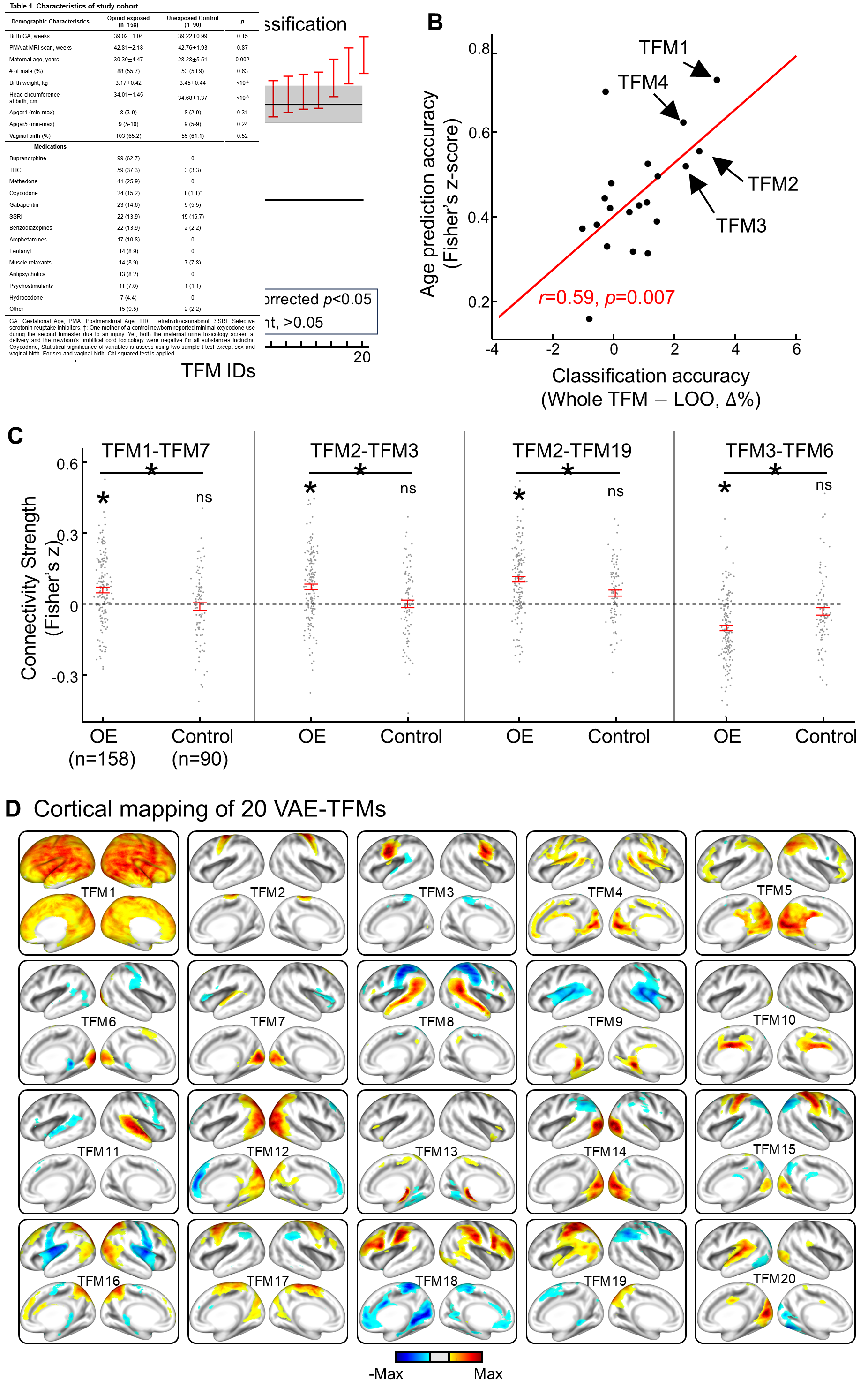 (A) Sensitivity analysis of individual TFMs to opioid exposure, assessed by excluding each TFM from the classification model. Red error bars indicate classification accuracy after exclusion; the gray shaded area represents baseline accuracy using all TFMs. (B) Relationship between each TFM's age-prediction performance and its sensitivity to opioid exposure, highlighting developmental relevance. (C) Connectivity between TFMs showing significant group differences. Each dot represents an individual subject; red error bars denote the standard error of the mean. OE: opioid-exposed group. * indicates Bonferroni-corrected p < 0.05 (two-sample t-test). (D) Full set of 20 temporal functional modes (TFMs) of healthy newborns derived using a variational autoencoder (VAE). TFMs defined in the VAE latent space are back-projected into a cortical space using the VAE decoder. The order of TFMs is set by its difference in classification accuracy between leave-one-TFM-out model vs. whole TFMs.