
Hematology/Oncology
Session: Hematology/Oncology 1
Shiyu Luo, PhD (she/her/hers)
Research assistant professor
University of Miami Leonard M. Miller School of Medicine
Miami, Florida, United States
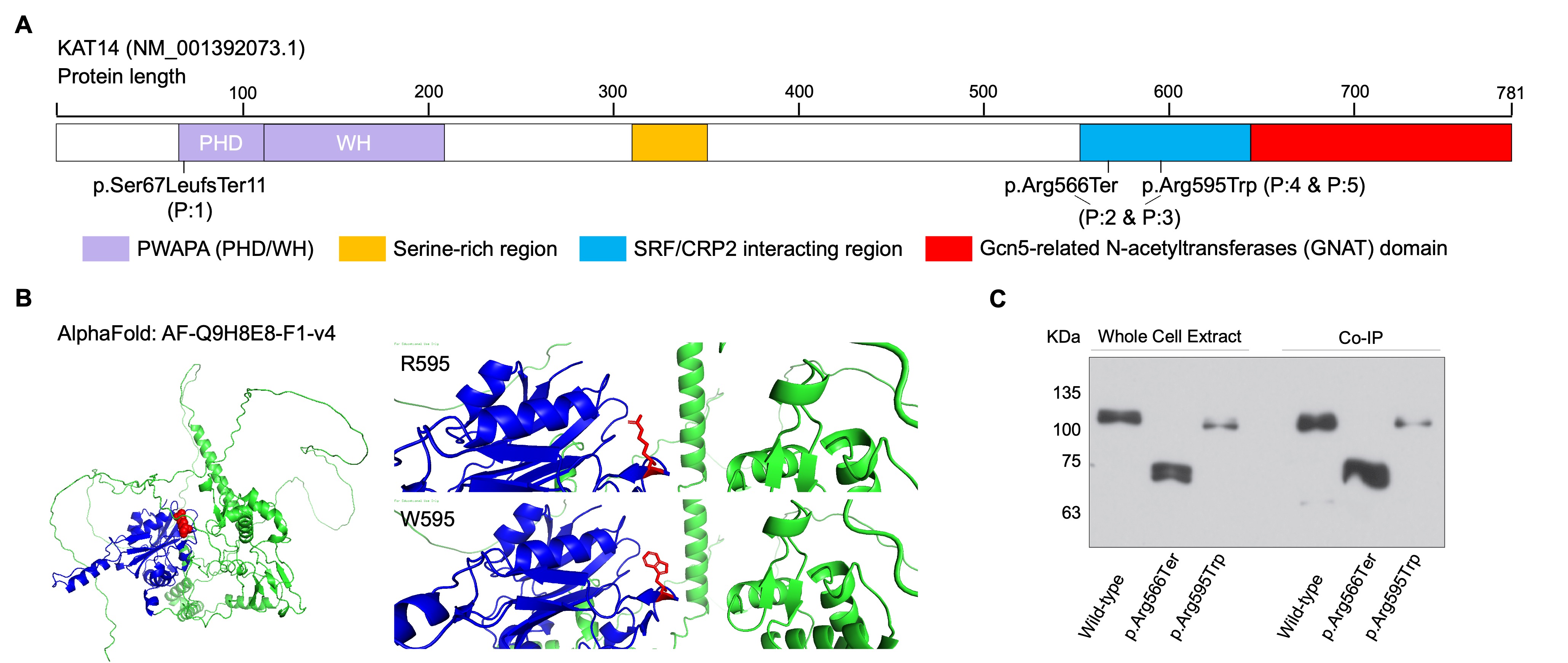 (A) Schematic representation of the human KAT14 gene (NM_001392073.1) highlighting the locations of three variants identified in patients: a frameshift (p.Ser67LeufsTer11), a nonsense (p.R566Ter), and a missense (p.R595W) variant. Prosite scan of the KAT14 protein sequence reveals a Gcn5-related N-acetyltransferases (GNAT)-type histone acetyltransferase (HAT) domain at the C-terminal region (amino acids 637 - 781). An interaction domain spanning residues 542-662 mediates binding to SRF and CRP2. Additional annotated features include a PWAPA cassette, comprising a PHD-like finger and a helix-winged-helix (WH) domain, potentially involved in chromatin targeting through recognition of histone modifications and DNA. A serine-rich region is located between amino acids 311-350. (B) Structural modeling of the p.R595W variant using the AlphaFold predicted structure (AF‑Q9H8E8‑F1‑v4) shows residue R596 (UniProt Q9H8E8 numbering) located on a surface-exposed helix at the periphery of the C-terminal lobe, just outside the GNAT domain. The substitution of arginine with tryptophan eliminates a positive charge and introduces a bulky aromatic side chain, predicted to disrupt local electrostatics and packing, potentially destabilizing the protein. Residues 573-782 are shown in blue, remainder in green. (C) Immunoblot analysis of KAT14 protein expression in HEK293 cells transfected with wild-type or variant constructs. As predicted, the p.Arg566Ter variant produced truncated proteins, while the p.Arg595Trp variant showed reduced steady-state protein levels, consistent with impaired stability. The p.Ser67LeufsTer11 variant identified in P:1 was not tested, as the patient was enrolled after completion of these cellular validation experiments.
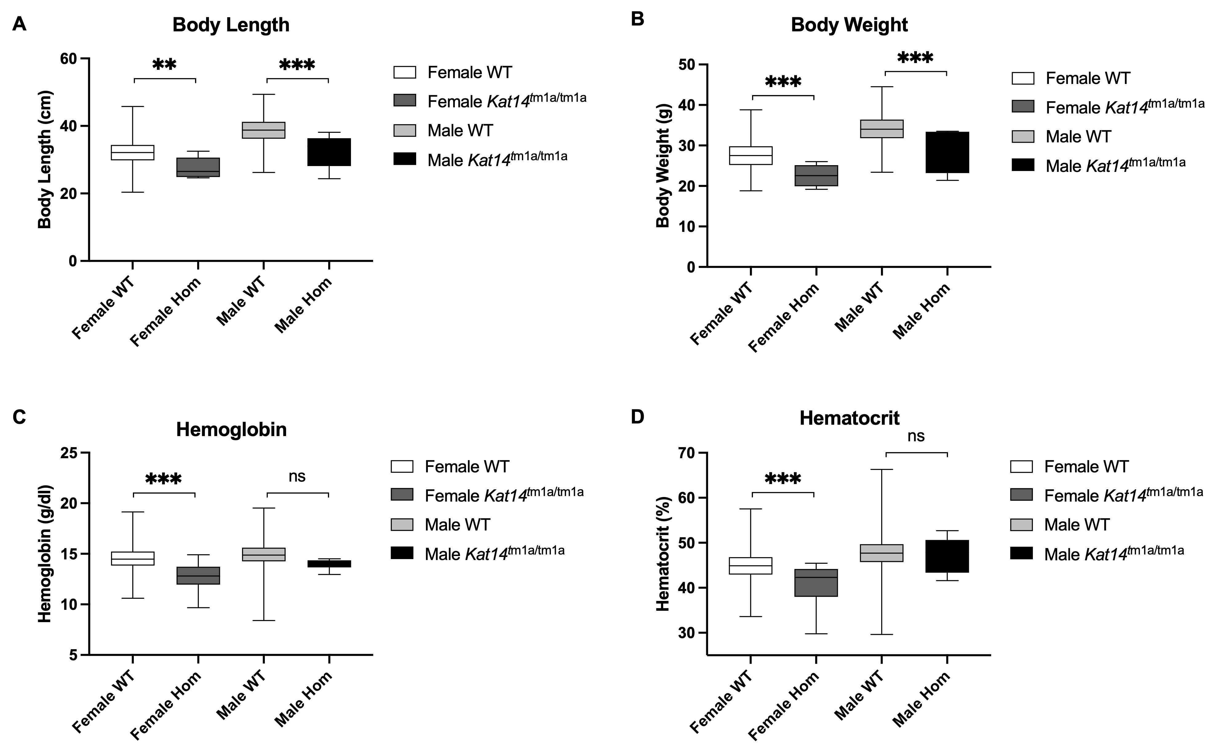 Comprehensive phenotyping data from the International Mouse Phenotyping Consortium (IMPC), generated through the Sanger Institute's Mouse Genetics Project (MGP), revealed that (A-B) Homozygous mutants show significantly decreased body length (A) and body weight (B) compared to wild-type controls. (C-D) Female homozygous mice exhibit hematological abnormalities including reduced hemoglobin concentration (C) and hematocrit (D). Data represent means ± SEM. Statistical significance was determined using unpaired t-tests. ** p < 0.01, *** p < 0.001.
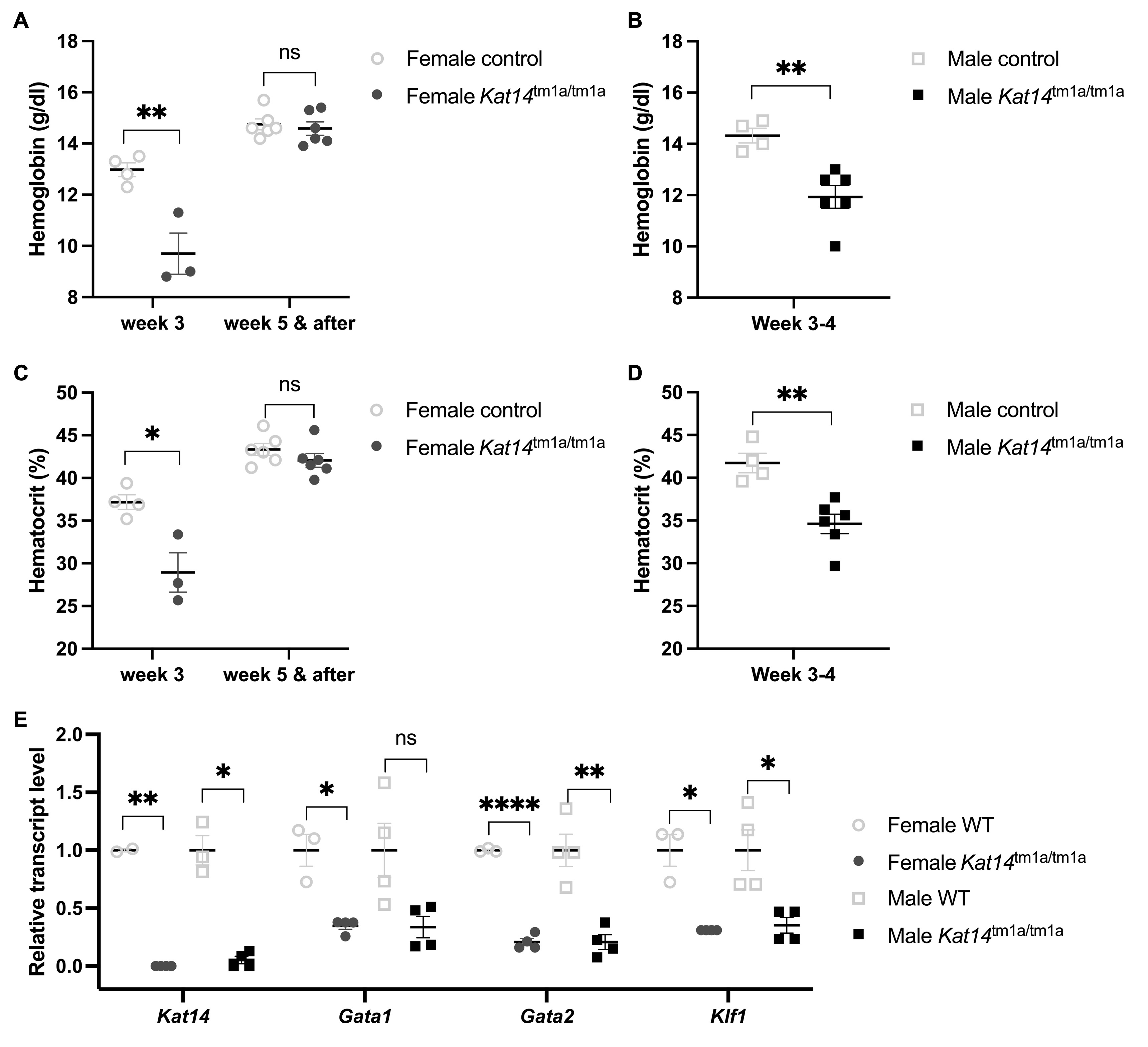 (A-D) Kat14tm1a/tm1a mice exhibited reduced hemoglobin (Hb; A, B) and hematocrit (Hct; C, D) at ~3 weeks of age, with normalization by 5 weeks. Female mice (A, C): n = 3 at 3 weeks of age, n = 6 at 5 weeks; and male mice (B, D): n = 4 at 3-4 weeks of age. (E) RT-qPCR analysis of erythroid-specific transcription factors Gata1, Gata2, and Klf1 using total RNA extracted from bone marrow of Kat14tm1a/tm1a mice at ~3 weeks of age. Gene expression was normalized to housekeeping controls and analyzed using the ΔΔCt method. Data represent mean ± SEM. Statistical comparisons were performed using unpaired t-tests or ANOVA as appropriate. *p < 0.05, **p < 0.01, ***p < 0.001, ****p < 0.0001.