601 - Social-Emotional Development and Functional Connectivity in Neonates
Sunday, April 26, 2026
9:30am - 11:30am ET
Publication Number: 3582.601
Caroline Wambach, Children's National Health System, Washington, DC, United States; Josepheen De Asis-Cruz, Children's National Health System, Bethesda, MD, United States; Julius Ngwa, Children's National Health System, Washington, DC, United States; Susan Weiner, Children's National Health System, Arlington, VA, United States; Catherine Limperopoulos, Children's National Health System, Washington DC, DC, United States; Nickie Andescavage, Children's National Health System, Washington, DC, United States
Fellow Children's National Health System Washington, District of Columbia, United States
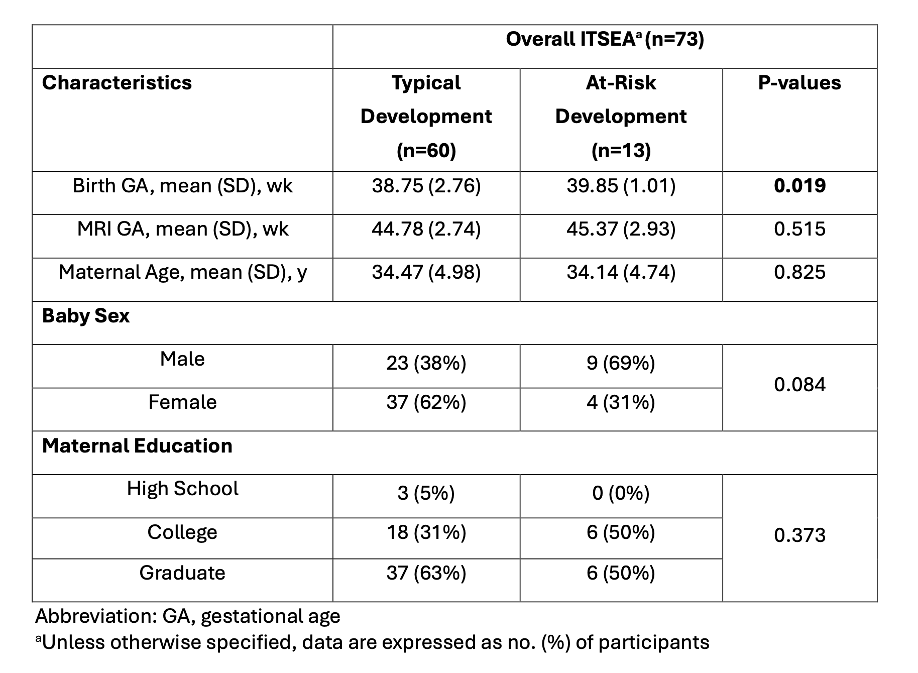
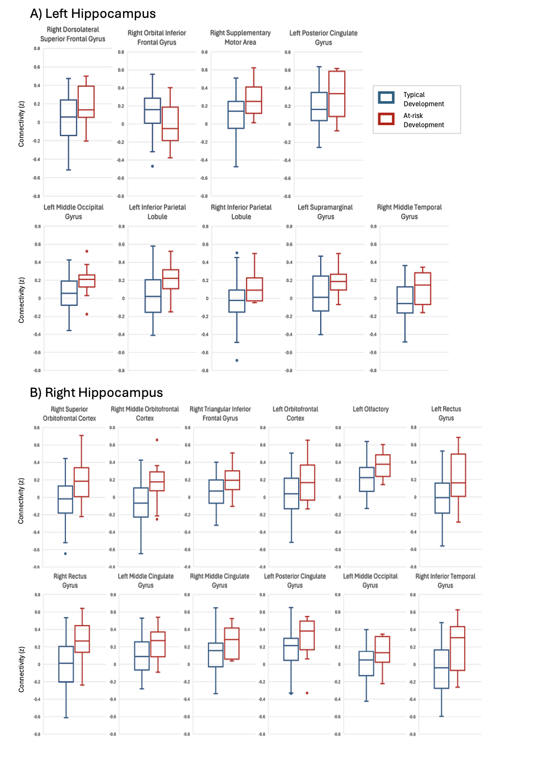
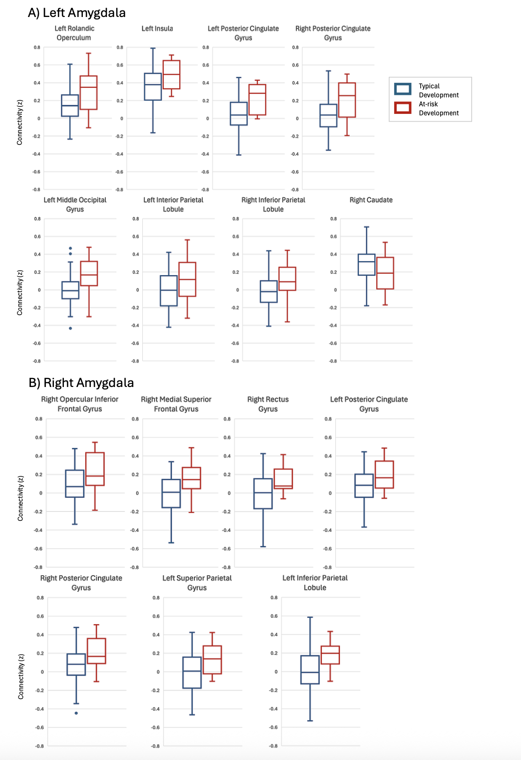
Background: The limbic system, including the amygdala (AMY) and hippocampus (HIP), is primarily responsible for processing emotions, memory, and motivation and therefore plays a critical role in social-emotional development. Prior neuroimaging research has shown that structural changes of the AMY and HIP secondary to stress exposure are associated with long-term neurodevelopment impairment including social-emotional problems. Functional connectivity (FC) of these limbic regions in relation to social-emotional development has not yet been explored. Objective: To evaluate the relationship between social-emotional development and FC of the AMY and HIP in neonates. Design/Methods: Mother-infant dyads were prospectively recruited into a longitudinal study. Neonatal resting-state functional MRI data were evaluated using a seed-based approach to assess FC of the bilateral HIP and AMY to 92 brain regions. Social-emotional development was assessed at 18 months using the Infant-Toddler Social-Emotional Assessment (ITSEA). Infants with a score indicating a deficit or delay on any of the ITSEA domains (externalizing, internalizing, dysregulation, and competence) were categorized as having an at-risk ITSEA. An ordinary least squares regression model was used to compare differences in FC in infants with and without social-emotional deficits. Results: A total of 73 mother-infant dyads were included in the analysis. There were no significant differences in baseline demographics except for gestational age at birth, which was adjusted for in the analyses. Overall, an at-risk ITSEA score was associated with relatively increased AMY/HIP FC to distributed ventral frontal, temporal, and cingulate regions encompassing the limbic and association cortices, and decreased FC between left HIP and right inferior orbital gyrus and left AMY and right caudate.
Conclusion(s): Our analysis suggests that social-emotional deficits may be associated with altered HIP/AMY FC. Toddlers with an at-risk ITSEA had a trend towards increased FC within limbic and association cortices, in line with prior research showing globally distributed hyperconnectivity in children with confirmed neurodevelopmental conditions. Although our findings did not survive multiple comparison correction, the direction of effects aligns with existing literature on emotional dysregulation and our prior research investigating maternal psychological distress and FC. Future studies are currently underway to assess whether these preliminary associations persist in larger samples and whether maternal psychological distress mediates the relationship between FC and social-emotional development.
Table 1: Demographic Characteristics by Overall ITSEA Score
Figure 1. Box plots demonstrating significant differences (p < 0.05) in functional connectivity of the (A) left hippocampus and (B) right hippocampus for typical vs at-risk development.
Figure 2. Box plots demonstrating significant differences (p < 0.05) in functional connectivity of the (A) left amygdala and (B) right amygdala for typical vs at-risk development.

 photo")