
Quality Improvement/Patient Safety
Session: Quality Improvement/Patient Safety 2
 photo")
Esra Abaci Turk, PhD (she/her/hers)
Assistant Professor of Pediatrics
Boston Children's Hospital, Harvard Medical School
Boston, Massachusetts, United States
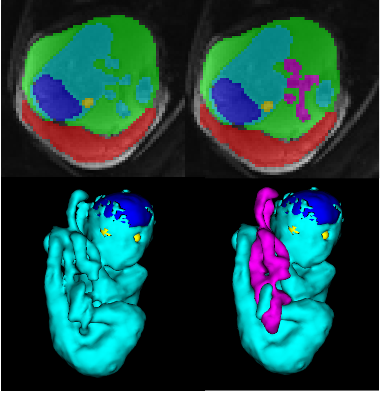 Whole uterus segmentation as they appear without and with the UC (manually segmented) in an example case from the training data of the UC segmentation model, in 2D (first row) and 3D (second row). Magenta: umbilical cord, cyan: fetal body, dark blue: fetal brain, yellow: fetal eyes, green: amniotic fluid, red: placenta.
 .png) Automatic (top row) and manual (bottom row) segmentations of the umbilical cord in 3D for the 4 test cases. Gestational ages are noted at the top and Dice scores are noted at the bottom for each test case. GA: gestational age, w: weeks, d: days.
Automatic (top row) and manual (bottom row) segmentations of the umbilical cord in 3D for the 4 test cases. Gestational ages are noted at the top and Dice scores are noted at the bottom for each test case. GA: gestational age, w: weeks, d: days. 