
Neonatal Neurology
Session: Neonatal Neurology 9: Pre-Clinical 1
Xiaodi F. Chen, MD, PhD (he/him/his)
Associate Professor
Women & Infants Hospital of Rhode Island
Providence, Rhode Island, United States
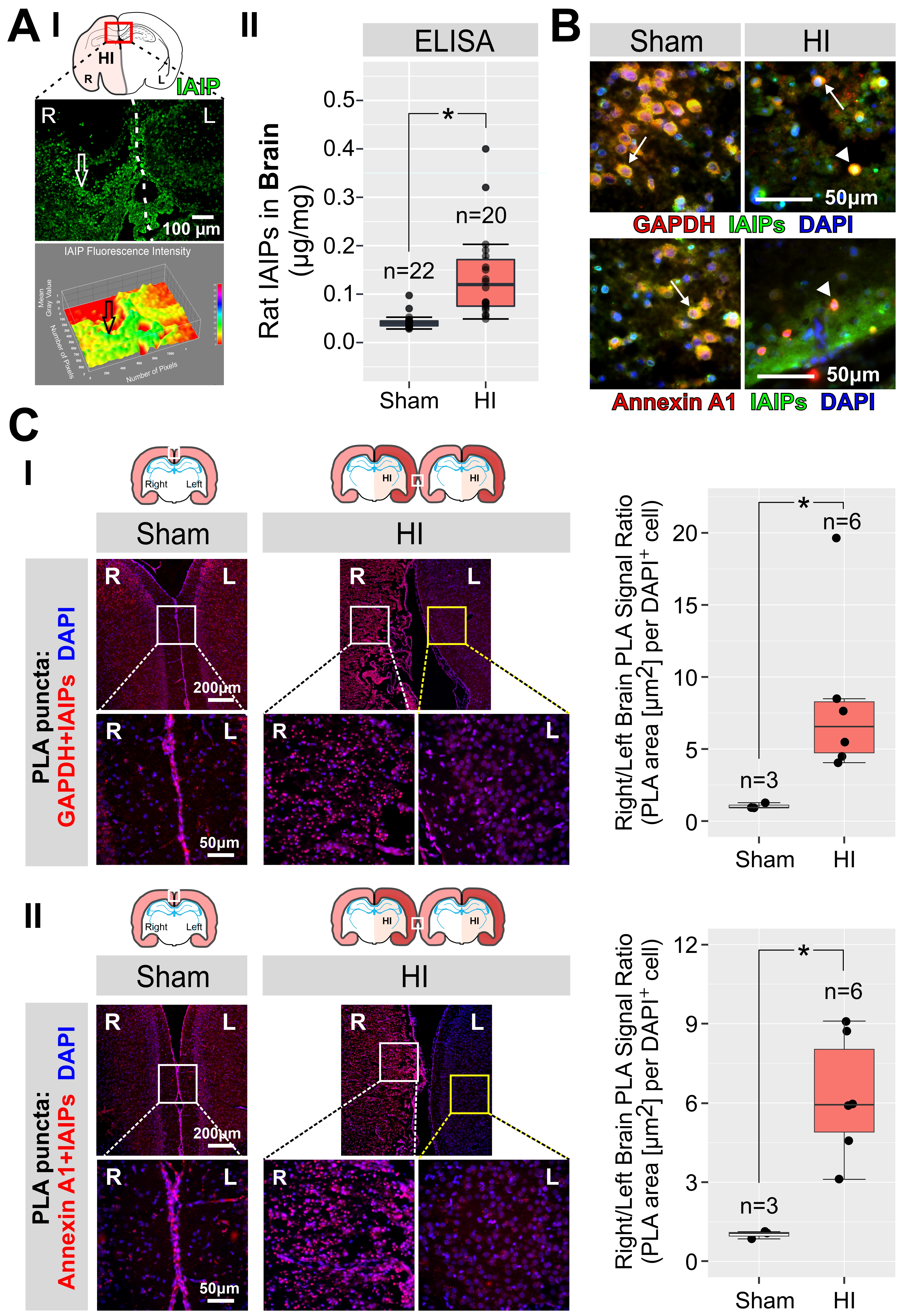 Figure A. IAIP expression in neonatal brain. (I) Representative immuno-fluorescence images show endogenous IAIP expression at P10 in the rat brain 72 h after exposure to HI. IAIP fluorescence intensity was higher in the ipsilateral (right) brain after HI compared with the contralateral non-ischemic (left) side (open arrows, top and bottom panels). (II) ELISA quantification demonstrates significant increases in IAIP levels 72 h after exposure to HI vs. Sham brains (*P <0.05). Statistical analysis: Mann-Whitney test.