297 - The Influence of Mesenchymal Stem Cells on the Severity of Hypox-ic-Ischemic Brain Damage in Neonatal Rats via Serum Cytokines
Monday, April 27, 2026
8:00am - 10:00am ET
Publication Number: 4292.297
Yuan Tan, The Fiest Affiliated Hospital of Guilin Medical University, Guilin, Guangxi, China (People's Republic); sifeng yue, The Fiest Affiliated Hospital of Guilin Medical University, Guilin, Guangxi, China (People's Republic); Feng Lin, Guilin Medical University, Chongqing, Chongqing, China (People's Republic); Zenghong Huang, THE FIRST AEEILATED HOSPITAL OF GECULLN MEDICAL UNIVERSITY, Guilin City, Guangxi, China (People's Republic); Xinyi Liang, 桂林医科大学第一附属医院, 桂林市, Guangxi, China (People's Republic); Shuchun Lyu, The first Affiliated hospital of Guilin medical university, Guilin, Guangxi, China (People's Republic)
Director of the Neonatal Department The Fiest Affiliated Hospital of Guilin Medical University Guilin, Guangxi, China (People's Republic)
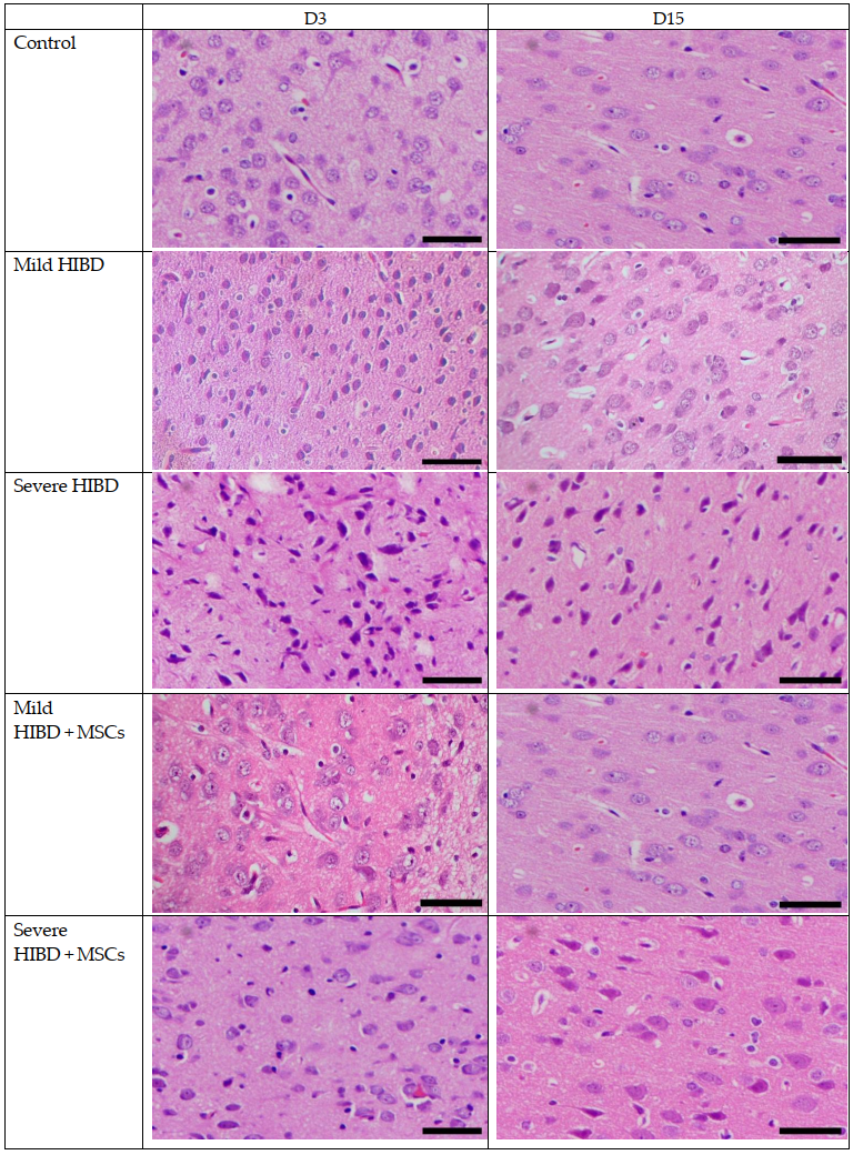
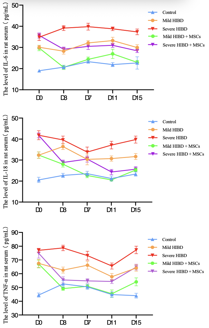
Background: Neonatal hypoxic-ischemic brain damage (Hypoxic‑Ischemic Brain Damage, HIBD) is a severe neurological disorder caused by perinatal asphyxia. Its severe long-term sequelae impose a heavy burden on families and society. Therefore, effective early diagnosis and intervention for HIBD are crucial for improving progno-sis. Objective: To investigate the correlation between serum levels of interleukin-6 (IL-6), interleukin-18 (IL-18), and tumor necrosis factor-α (TNF-α) and the severity of hypoxic-ischemic brain damage (HIBD) in neonatal SD rats, as well as the effects of mesenchymal stem cells (MSCs) on serum cytokine levels and the repair of HIBD in rats. Design/Methods: 250 7-day-old SD rats were selected and a HIBD model was established using the Rice-Vannucci method. The rats were randomly divided into 5 groups: 1) control group; 2) mild HIBD group; 3) severe HIBD group; 4) mild HIBD + MSCs group; 5) severe HIBD + MSCs group. The MSCs intervention group rats were injected with MSCs into the lumbar spine 1 and 8 days after modeling. Longa scores were evaluated at 0, 3, 7, 11, and 15 days after modeling, and serum levels of IL-6, IL-18, and TNF-α were detected. Brain tissue was stained with hematoxylin-eosin (HE) for Longa scoring. The correlation between serum cytokines and the severity of brain injury was analyzed, and the regulatory effect of MSCs intervention on cytokine expression levels and its role in neural injury repair were further explored. Results: (1) Both mild and severe HIBD groups exhibited significantly higher Longa scores and serum IL-6, IL-18, and TNF-α levels than the control group at all stages (P < 0.05), with the severe group showing further elevation over the mild group (P < 0.05). (2) Post-treatment HE staining indicated improved brain tissue pathology in the mild HIBD and mild HIBD + MSCs groups. Significant reductions in Longa scores and cytokine levels were observed at all post-treatment time points (D3, D7, D11, D15) compared to baseline (D0) in the mild HIBD, mild HIBD + MSCs, and severe HIBD + MSCs groups.
Conclusion(s): The serum levels of IL-6, IL-18, and TNF-α in rats are positively correlated with the severity of HIBD and can reflect the degree of brain tissue injury. They can be used for disease assessment; mesenchymal stem cell treatment can significantly reduce the serum levels of IL-6, IL-18, and TNF-α in HIBD rats, alleviate in-flammatory responses, improve symptoms, and repair brain injury.
Figure 1. HE staining of paraffin section of brain tissue in SD rats (scale bar :50μm)
Figure 2、3、4 The level of IL-6/IL-18/TNF-α in rat serum