
Neonatal Fetal Nutrition & Metabolism
Session: Neonatal Fetal Nutrition & Metabolism 2
Liliette I. Quintana, B.A. (she/her/hers)
Research Technician
Weill Cornell Medicine
New York, New York, United States
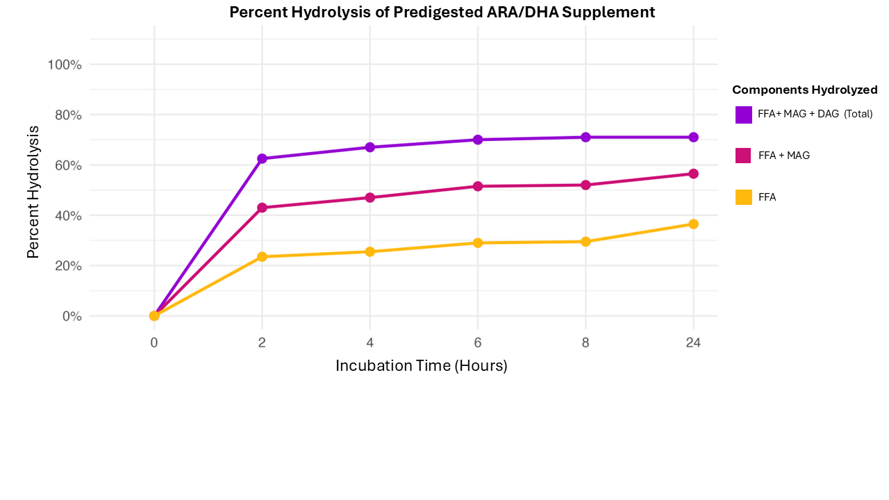 Figure 1: Modeling of Predigestion System Prior to in vitro Experiments. Hydrolysis confirmed by bands of Free Fatty Acids (FFA), Monoglycerides (MAG), Diglycerides (DAG) over all components and Triglyceride Base (TAG). Hydrolysis determined by Thin Layer Chromatograph (TLC), timepoints between 0-24 Hours.
 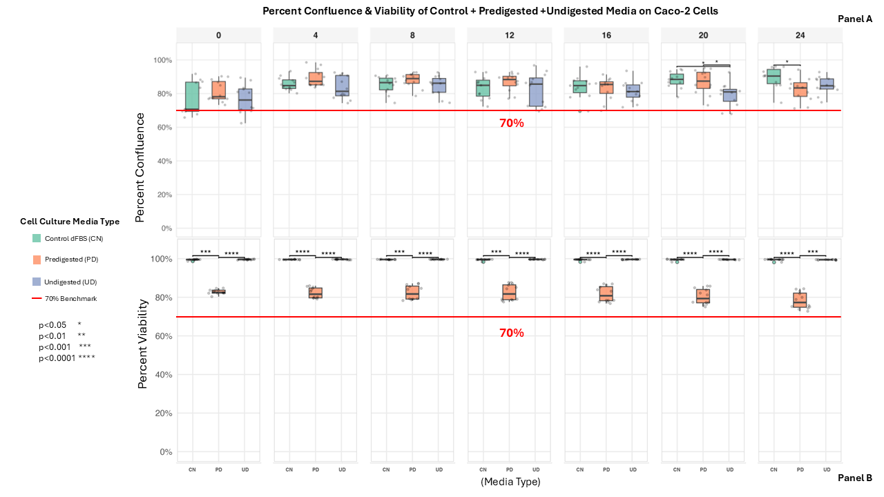 Figure 2: Threshold for Predigestion Cell Culture Experiments. Caco-2 cells were incubated with CN, PD, and UD media to assess whether ≥70% confluence (Panel A) and viability (Panel B) could be maintained for downstream stress assays. Box-and-whisker plots demonstrate that all groups remained above the 70% threshold, confirming suitability for subsequent experiments.
.png) Figure 3. Wound Gap Closure in Scratch Assay. Panel A: Box and whisker plot of relative wound closure with control and predigested/undigested meal in MEM media during scratch assay. Caco-2 cells exposed to various media from timepoints between 0-24h. Panel B: Cellular recovery after scratch-wound was visible through wound closure in 10x wide field imaging of Caco-2 cells in dFBS control (CN), Predigested (PD) and Undigested (UD). Blue Mask indicates wound, yellow mask indicates epithelial cell growth after initial scratch.
Figure 3. Wound Gap Closure in Scratch Assay. Panel A: Box and whisker plot of relative wound closure with control and predigested/undigested meal in MEM media during scratch assay. Caco-2 cells exposed to various media from timepoints between 0-24h. Panel B: Cellular recovery after scratch-wound was visible through wound closure in 10x wide field imaging of Caco-2 cells in dFBS control (CN), Predigested (PD) and Undigested (UD). Blue Mask indicates wound, yellow mask indicates epithelial cell growth after initial scratch.