
Neonatal Neurology
Session: Neonatal Neurology 5: Preterm & Neurodevelopment
 photo")
Hideyoshi Fukui, MD (he/him/his)
PhD Candidate, Neonatologist
Department of neonatology, Izumiotsu Women's and Children's Hospital, Osaka Metropolitan University Graduate School of Medicine
Izumiotsu-shi, Osaka, Japan
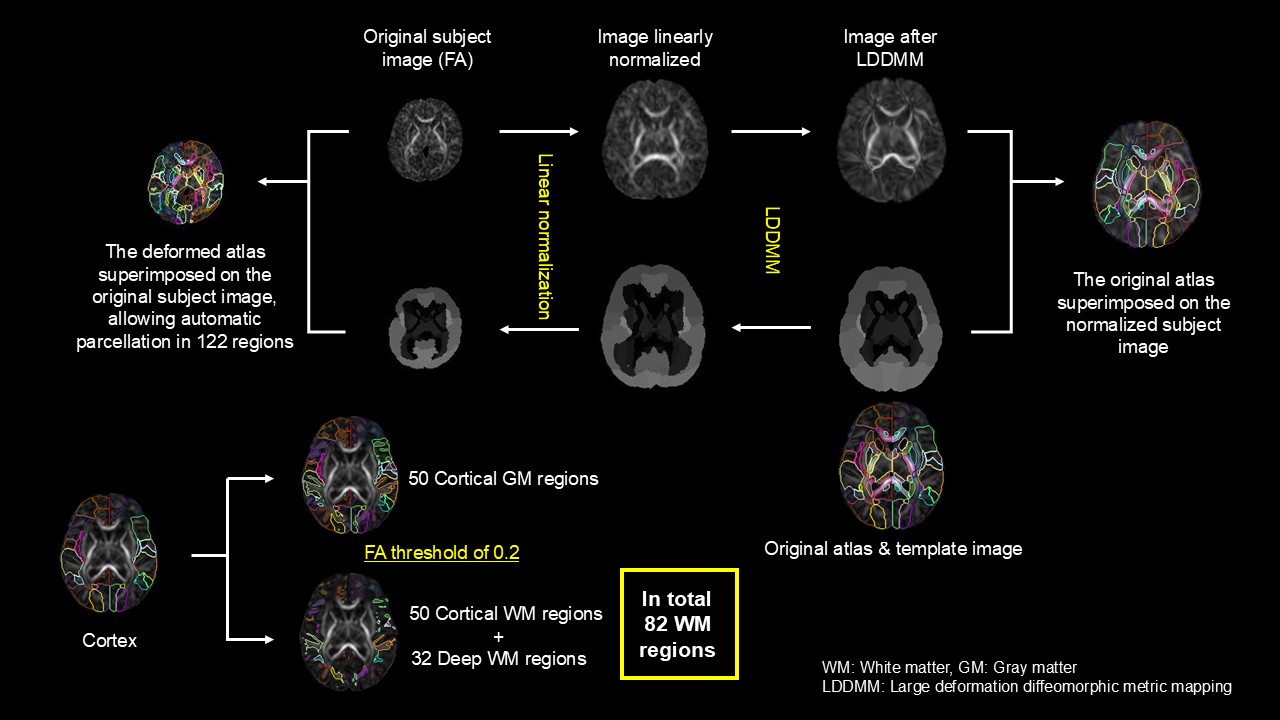 DiffeoMap (Johns Hopkins University) was used to first linear normalize each subject FA and b0 brain images to the template FA and b0 common space, respectively, followed by a non-rigid registration (Large deformation diffeomorphic metric mapping, LDDMM) to obtain the registration matrices. Then, neonatal brain atlas (Oishi, 2011) was transformed to fit each subject brain by inverse registration (LDDMM followed by inverse linear registration) using the matrices obtained, and superimposed on the original subject image, allowing parcellation of 122 regions. Fifty cortical regions were then separated into 50 cortical GM and WM regions by threshold of FA 0.2, ending up with automatically parcellating in total 82 WM regions from the whole brain.
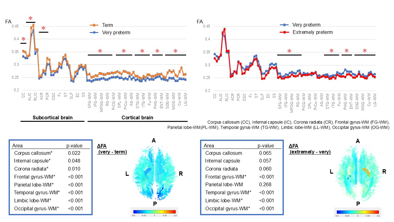 Very preterm infants showed significantly lower FA values in corpus callosum (CC), internal capsule (IC), corona radiata (CR) and all cortical WM compared with term infants. Extremely preterm infants showed significantly lower FA values in all cortical WM except for parietal region, compared with very preterm infants.
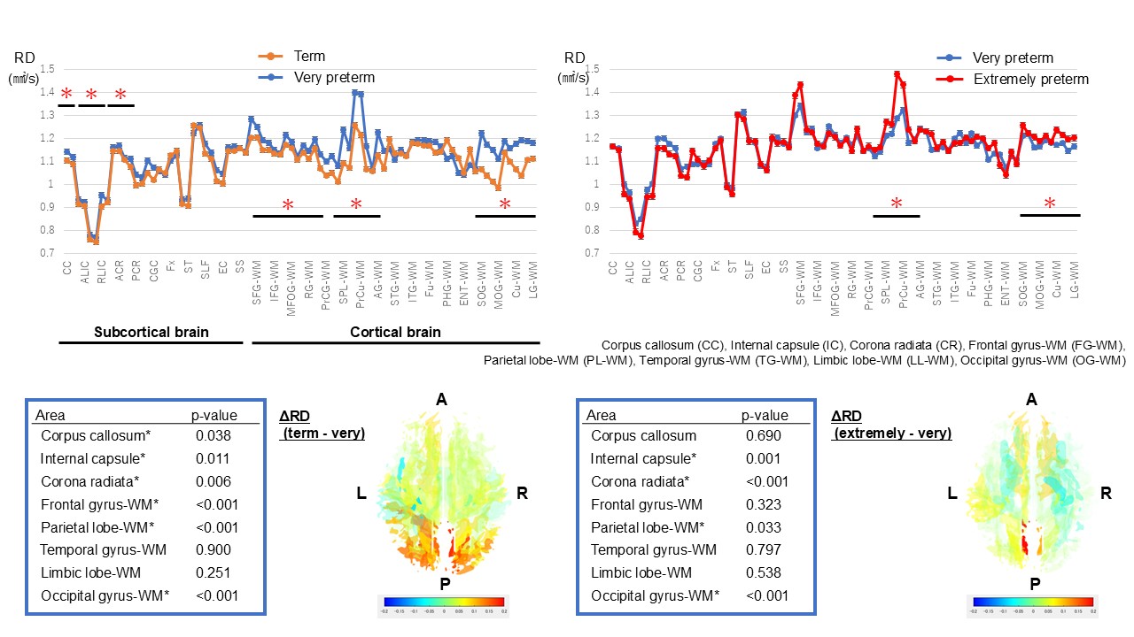 Very preterm infants showed significantly higher RD in CC, IC, CR and FG-, PL- and OG-WM compared with term infants. Extremely preterm infants showed significantly higher RD in PL- and OG-WM compared with very preterm infants.