504 - A Novel Artificial Intelligence (AI) Workflow for 3D Glomerular Reconstruction and Quantification from Routine Serial Sections
Saturday, April 25, 2026
3:30pm - 5:45pm ET
Publication Number: 2490.504
Andrew Rauch, Monroe Carell Jr. Children's Hospital at Vanderbilt, Nashville, TN, United States; Yuqing Liu, Department of Nephrology, Shanghai Tongji Hospital, School of Medicine, Tongji University, Shanghai, Shanghai, China (People's Republic); Yanfan Zhu, Vanderbilt University, Nashville, TN, United States; Jianyong Zhong, Vanderbilt University School of Medicine, Nashville, TN, United States; Valentina Kon, Vanderbilt University School of Medicine, Nashville, TN, United States; Yuankai Huo, Vanderbilt University, Nashville, TN, United States; Haichun Yang, Vanderbilt University Medical Center, Nashville, TN, United States
Pediatric Nephrology Fellow Monroe Carell Jr. Children's Hospital at Vanderbilt Nashville, Tennessee, United States
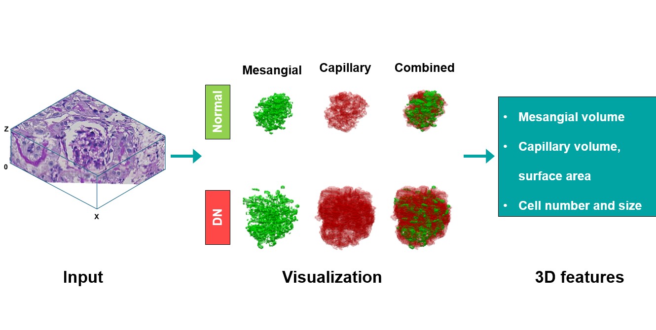
Background: Pediatric kidney disease is a significant global health burden that often requires a kidney biopsy for diagnosis. A major feature in these renal biopsies is glomerular pathology. Currently, pathological diagnosis relies on 2D images and morphological features viewed on one slide at a time, even though serial sections are routinely available. This conventional approach overlooks potentially critical structural information that can be gleaned from 3D images. Objective: To establish an AI-based workflow for 3D reconstruction and quantification of glomeruli from standard serial sections. Demonstrate utility of the workflow by exposing intricate morphological features in a diabetic nephropathy (DN) model. Design/Methods: We developed an integrated AI pipeline. Paraffin blocks from normal and diabetic nephropathy (DN) mice (db/db eNOS KO, 10 weeks) were serially sectioned (50 slices) and Periodic Acid-Schiff (PAS)-stained. Following expert annotation of the glomerular tuft, mesangium, and cells, our AI algorithm achieved a robust automated alignment of all the serial cross-sections, 3D reconstruction, and quantitative feature extraction. Results: The workflow successfully generated precise 3D glomerular models (see figure). Application to the model quantified disease-specific 3D features inaccessible to routine 2D assessment. In glomeruli of DN kidneys, AI calculated a 1.25-fold increase in the glomerular tuft volume, a 1.74-fold increase in the mesangial volume, and a 3.33-fold increase in glomerular capillary volume vs control glomeruli. The capillary surface area increased 1.78-fold. DN glomeruli had more than double the cell count (360 vs. 154/glomerulus) despite a smaller size of individual cells (69.8 vs. 86.6 µm³).
Conclusion(s): We have established a unique AI workflow that transforms routine glomerular serial sections into quantifiable 3D glomerular models. This tool presents a novel, objective 3D metric that demonstrates significant potential to enhance the accuracy and depth of pathological evaluation of kidney biopsies that can provide standardization in diagnostic criteria to predict disease progression, enable personalized treatment strategies, and enhance outcomes in children with kidney disease.
3D Glomerular Reconstruction: Normal vs. Diabetic Nephropathy (DN)

.jpg "Andrew Rauch, DO photo")