
Neonatal Neurology
Session: Neonatal Neurology: Pre-Clinical Research Trainee Ongoing Projects
 photo")
Stephanie Newman, MD (she/her/hers)
Resident Physician
Children's Hospital of The King's Daughters
Norfolk, Virginia, United States
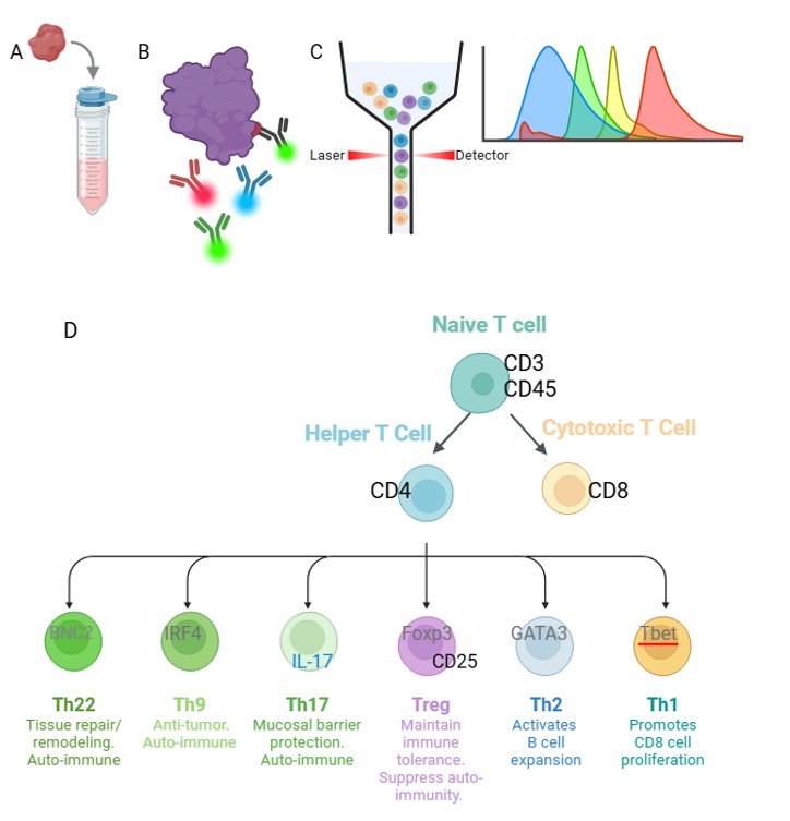 A) Mice were humanely euthanized using lethal injection of phenobarbital followed by immediate harvesting of tissues. Tissues were passed through 70um filter with ACK lysis buffer to isolate lymphocytes. B) Cell suspensions were incubated with a cocktail of fluorescent-conjugated antibodies to stain for surface and intracellular proteins for identification and activation status. C) Samples were quantified via Cytek Aurora Spectral Flow Cytometer and data analyzed in FlowJo. D) Panel of surface (black), intracellular (blue), and nuclear (gray) markers used to identify and quantify activation status of lymphocyte subsets.
 .jpg) Cerebral levels of T lymphocytes, both CD4+ and CD8+, increased in HI-injured rats (NT and TH) from 3 to 11 dpi (A-C). Similarly, levels of transcription factors for Th1 (Tbet), Th2 (GATA3), Treg (Foxp3) and Th17 (IL17a) differentiation all increased in NT and TH but not Sham from 3 to 11 dpi. (D-G) Graphs display means and standard error. N=6 rats per treatment group per harvest point.
Cerebral levels of T lymphocytes, both CD4+ and CD8+, increased in HI-injured rats (NT and TH) from 3 to 11 dpi (A-C). Similarly, levels of transcription factors for Th1 (Tbet), Th2 (GATA3), Treg (Foxp3) and Th17 (IL17a) differentiation all increased in NT and TH but not Sham from 3 to 11 dpi. (D-G) Graphs display means and standard error. N=6 rats per treatment group per harvest point. .jpg) Cerebral levels of T helper cells (A) and Treg cells (B) increased in over time following injury, and both were higher in injured female rats than male rats. Graphs show means and standard error. *indicates t test p-values <0.05. N=6 rats per treatment group per day, with 6 males and females for each sex.
Cerebral levels of T helper cells (A) and Treg cells (B) increased in over time following injury, and both were higher in injured female rats than male rats. Graphs show means and standard error. *indicates t test p-values <0.05. N=6 rats per treatment group per day, with 6 males and females for each sex.