Session: Neonatal Neurology: Pre-Clinical Research Trainee Ongoing Projects
TOP 78 - Investigating the ontogeny of microglial migration into the fetal hippocampus in a mouse model of IUGR
Sunday, April 26, 2026
9:30am - 11:30am ET
Publication Number: 3810.TOP 78
Frank A. Strnad, University of Utah School of Medicine, South Salt Lake, UT, United States; Ashley Brown, University of utah, Salt lake, UT, United States; Matthew Wieben, University of Utah, Cottonwood Heights, UT, United States; Camille M. Fung, University of Utah, Dept. of Pediatrics, Salt Lake City, UT, United States
Fellow University of Utah School of Medicine South Salt Lake, Utah, United States
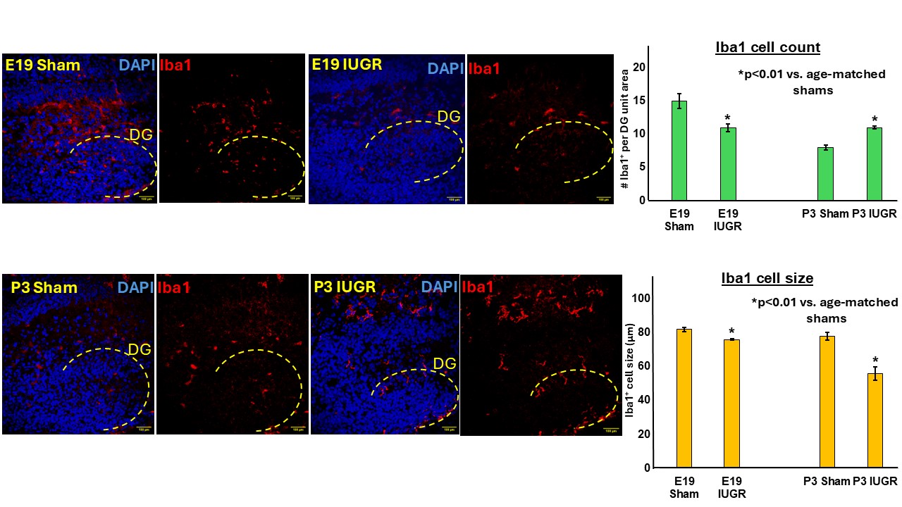
Background: Hypertensive diseases of pregnancy (HDP) causing uteroplacental insufficiency (UPI) is the most common cause of intrauterine growth restriction (IUGR) in developed countries. IUGR offspring are at 5x increased risks for learning and memory deficits. We have previously published that the IUGR hippocampal dentate gyrus (DG) neurons have decreased dendritic branching and volume one month after birth. This is seen in association with increased microglial number and cell size potentially pruning the dendrites. We know that microglia migrate into the hippocampus from the fetal yolk sac, are then amplified first in the aorta-gonad-mesonephros (AGM) region and then in the liver and eventually enter the brain via the choroid plexus in the lateral ventricles (LV). Whether UPI promotes embryonic microglial migration into the hippocampus to result in increased microglial number is unknown. We hypothesize that UPI recruits more DG microglia with increased cell size prenatally that persist into postnatal life. Objective: We aim to investigate how UPI affects the ontogeny of microglia migration into the DG in a translational mouse model of HDP/UPI/IUGR at embryonic day (E) 19 and postnatal day (P)3. Design/Methods: We are using our laboratory's mouse model of HDP/UPI/IUGR by implanting a micro-osmotic pump containing either vehicle (sham) or a potent vasoconstrictor TXA2-analog in C57BL/6 pregnant dams at E12.5 (term gestation ~20 days). We harvested brains at E19 and P3. We performed immunofluorescent staining for Iba1+ microglia and DAPI for cell nuclei. We have captured DG and LV images at 40x magnification. We are counting Iba1+ microglia number and measuring Iba1+ cell size using NIH ImageJ software. We will perform a two-way ANOVA test to compare the means of treatment (sham or IUGR) and sex (male or female) for each age and at each location imaged. Using a priori power analysis with two-sided testing, an effect size 1.6, α error probability 0.05, power 0.8, and allocation ratio N2/N1, we expected a sample size of 8 per sham, IUGR, males or females. We anticipate that all our measurements will be completed by Jan 31, 2026.
Figure 1. Representative immunohistochemical images for microglia staining and quantitative bar graph of Iba1 cell count and size in E19 and P3 hippocampal dentate gyrus.

 photo")