
Quality Improvement/Patient Safety
Session: Quality Improvement/Patient Safety 3
 photo")
Filiz Yetisir, PhD (she/her/hers)
Instructor
Harvard Medical School
Boston, Massachusetts, United States
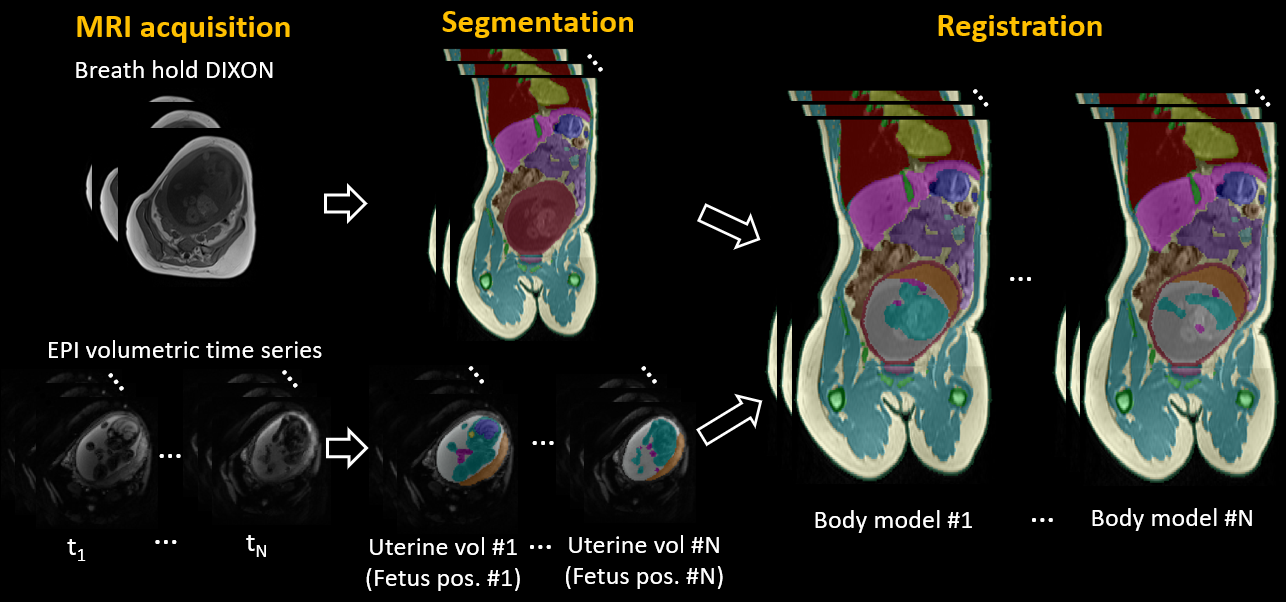 Pipeline to generate several body models with different fetal positions that sample fetal motion from one pregnant subject.
 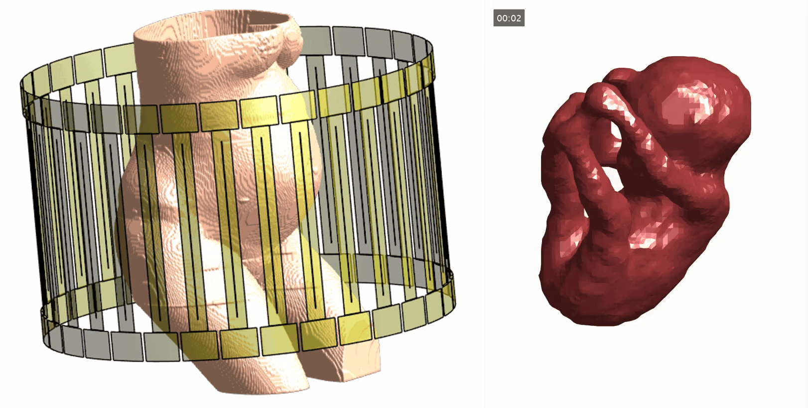 Left: maternal body model loaded into the RF coil model, right: dynamic fetus model. Fetal motion was observed and sampled during ~15 mins of EPI acquisition, however the effective fetal motion duration shown is ~10 mins after the elimination of fetal positions with severe motion artifacts or poor segmentation quality. Time stamp shows the time for 270 fetal positions with a sampling interval of 2.1 s, not the actual time of each fetal position.
.png) Normalized local SAR maps through peak maternal SAR (left) and peak fetal SAR for example fetal positions. Black plus signs show the peak maternal or fetal SAR location.
Normalized local SAR maps through peak maternal SAR (left) and peak fetal SAR for example fetal positions. Black plus signs show the peak maternal or fetal SAR location. 