289 - Postnatal environmental enrichment improves hippocampal dentate gyrus dendritic morphology and memory function in juvenile IUGR mouse offspring
Monday, April 27, 2026
8:00am - 10:00am ET
Publication Number: 4284.289
Camille M. Fung, University of Utah, Dept. of Pediatrics, Salt Lake City, UT, United States; Ashley S. Alvarado, University of Utah, Salt Lake City, UT, United States; Matthew Wieben, University of Utah, Cottonwood Heights, UT, United States; Frank Strnad, University of Utah School of Medicine, Salt Lake City, UT, United States; Megan Williams, University of Utah School of Medicine, salt lake city, UT, United States
Associate Professor University of Utah, Dept. of Pediatrics Salt Lake City, Utah, United States
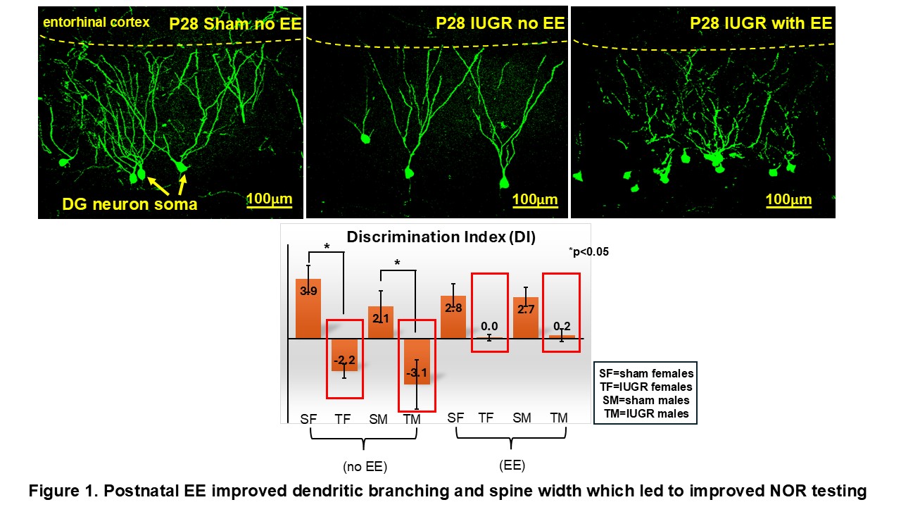
Background: Offspring born with intrauterine growth restriction (IUGR) have a 5-fold higher risk for learning and memory impairment compared to appropriately-grown (AG). In developed countries, hypertensive disorders of pregnancy (HDP) cause uteroplacental insufficiency (UPI) leading to IUGR. We have previously shown in a translational mouse model of HDP/UPI that IUGR hippocampal dentate gyrus (HDG) has reduced dendritic branching and volume at a month of age that leads to adult memory deficits. Objective: We tested whether postnatal environmental enrichment (EE) in juvenile mice, performed during a period of synaptic plasticity, could improve hippocampal DG dendritic morphology and memory deficit. Design/Methods: We replicated HDP/UPI and produced IUGR mice by a continuous micro-osmotic pump infusion of a potent vasoconstrictor, TXA2-analog, in timed-pregnant C57BL/6 mouse dams beginning on embryonic day (E)12.5 until term (~20 days). Sham-operated dams with vehicle infusion produced AG mice. After delivery, mice were cross-fostered and raised to postnatal day (P) 21. We injected a recombinant adeno-associated virus conjugated to green fluorescent protein to label DG dendritic spines. We placed half of P21 sham and IUGR offspring into postnatal EE for one hour each day for 7 days, while the other half remained in their home cages. Postnatal EE mimics a playground with colorful toys to enhance sensory, motor, social, and cognitive stimuli. At P28, we harvested brains for immunofluorescent staining of GFP-labeled DG neurons. At P60, we performed novel object recognition (NOR) for memory testing. Results: Juvenile IUGR DG dendrites had no change in spine width before or after EE. In contrast, they had increased spine density (= total spine count/dendritic length; 0.137±0.014 IUGR vs. 0.111±0.010 in AG, p< 0.05) compared to AG before EE. Postnatal EE normalized this spine density difference between AG and IUGR. Most interestingly, IUGR mice showed a baseline negative discrimination index (DI) compared to AG mice in NOR, but EE reversed it towards a positive index, showing that IUGR mice preferentially explored the novel object on next day similar to AG mice (Fig.1).
Conclusion(s): Postnatal EE, performed during a critical period of synaptic plasticity in juvenile IUGR offspring, could improve hippocampal neuronal morphology and function. This non-invasive therapy is reminiscent of Early Intervention, which should be prescribed to infants with IUGR to improve their lifelong risk for memory impairment. Unless an infant is born extremely preterm or has ELBW, doing nothing is the current standard of care for infants with IUGR.
Figure 1. Postnatal EE improved dendritic branching and spine width which led to improved NOR testing in IUGR offspring

 photo")